


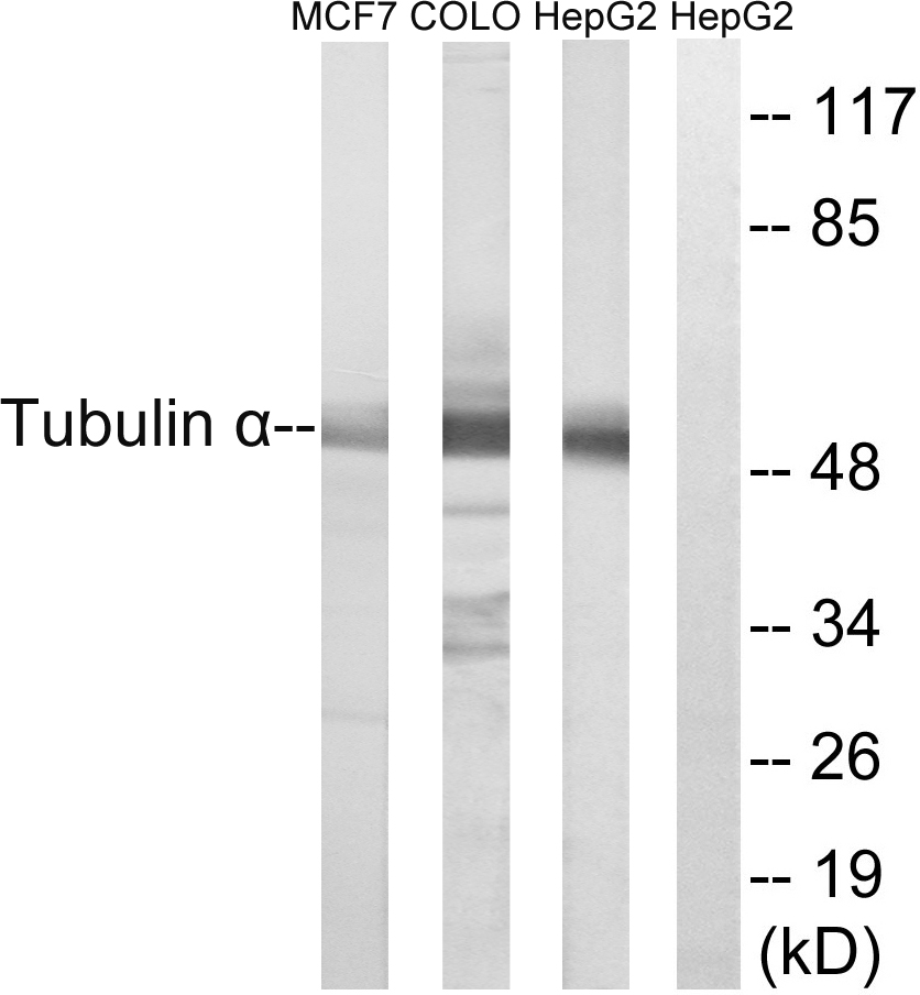
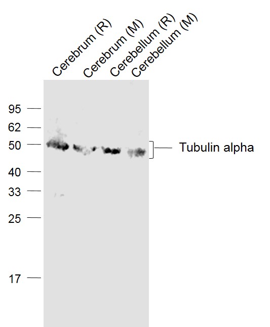
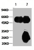
Figure 1. Western blot analysis of Tubulin alpha using anti-Tubulin alpha antibody (A03989-1). Electrophoresis was performed on a 5-20% SDS-PAGE gel at 70V (Stacking gel) / 90V (Resolving gel) for 2-3 hours. The sample well of each lane was loaded with 30 ug of sample under reducing conditions. Lane 1: human Hela whole cell lysates, Lane 2: human THP-1 whole cell lysates, Lane 3: human HEL whole cell lysates, Lane 4: human HepG2 whole cell lysates, Lane 5: rat brain tissue lysates, Lane 6: rat testis tissue lysates, Lane 7: mouse brain tissue lysates, Lane 8: mouse testis tissue lysates. After Electrophoresis, proteins were transferred to a Nitrocellulose membrane at 150mA for 50-90 minutes. Blocked the membrane with 5% Non-fat Milk/ TBS for 1.5 hour at RT. The membrane was incubated with rabbit anti-Tubulin alpha antigen affinity purified polyclonal antibody (Catalog # A03989-1) at 0.25 microg/mL overnight at 4°C, then washed with TBS-0.1%Tween 3 times with 5 minutes each and probed with a goat anti-rabbit IgG-HRP secondary antibody at a dilution of 1:10000 for 1.5 hour at RT. The signal is developed using an Enhanced Chemiluminescent detection (ECL) kit (Catalog # EK1002) with Tanon 5200 system. A specific band was detected for Tubulin alpha at approximately 50 kDa. The expected band size for Tubulin alpha is at 50 kDa.
. Tubulin alpha was detected in paraffin-embedded section of human rectum adenocarcinoma tissue. Heat mediated antigen retrieval was performed in EDTA buffer (pH8.0, epitope retrieval solution). The tissue section was blocked with 10% goat serum. The tissue section was then incubated with 2microg/ml rabbit anti-Tubulin alpha Antibody (A03989-1) overnight at 4°C. Peroxidase Conjugated Goat Anti-rabbit IgG was used as secondary antibody and incubated for 30 minutes at 37°C. The tissue section was developed using HRP Conjugated Rabbit IgG Super Vision Assay Kit (Catalog # SV0002) with DAB as the chromogen.")
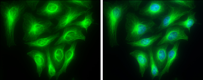
. Tubulin alpha was detected in immunocytochemical section of A549 cells. Enzyme antigen retrieval was performed using IHC enzyme antigen retrieval reagent (AR0022) for 15 mins. The cells were blocked with 10% goat serum. And then incubated with 5microg/mL rabbit anti-Tubulin alpha Antibody (A03989-1) overnight at 4°C. DyLight®488 Conjugated Goat Anti-Rabbit IgG (BA1127) was used as secondary antibody at 1:100 dilution and incubated for 30 minutes at 37°C. The section was counterstained with DAPI. Visualize using a fluorescence microscope and filter sets appropriate for the label used.")
. Tubulin alpha was detected in immunocytochemical section of NRK cells. Enzyme antigen retrieval was performed using IHC enzyme antigen retrieval reagent (AR0022) for 15 mins. The cells were blocked with 10% goat serum. And then incubated with 5microg/mL rabbit anti-Tubulin alpha Antibody (A03989-1) overnight at 4°C. DyLight®550 Conjugated Goat Anti-Rabbit IgG (BA1135) was used as secondary antibody at 1:100 dilution and incubated for 30 minutes at 37°C. The section was counterstained with DAPI. Visualize using a fluorescence microscope and filter sets appropriate for the label used.")
. Overlay histogram showing Hela cells stained with A03989-1 (Blue line). To facilitate intracellular staining, cells were fixed with 4% paraformaldehyde and permeabilized with permeabilization buffer. The cells were blocked with 10% normal goat serum. And then incubated with rabbit anti-Tubulin alpha Antibody (A03989-1,1microg/1x106 cells) for 30 min at 20°C. DyLight®488 conjugated goat anti-rabbit IgG (BA1127, 5-10microg/1x106 cells) was used as secondary antibody for 30 minutes at 20°C. Isotype control antibody (Green line) was rabbit IgG (1microg/1x106) used under the same conditions. Unlabelled sample without incubation with primary antibody and secondary antibody (Red line) was used as a blank control.")
Figure 1. Western blot analysis of Tubulin alpha using anti-Tubulin alpha antibody (A03989-1). Electrophoresis was performed on a 5-20% SDS-PAGE gel at 70V (Stacking gel) / 90V (Resolving gel) for 2-3 hours. The sample well of each lane was loaded with 30 ug of sample under reducing conditions. Lane 1: human Hela whole cell lysates, Lane 2: human THP-1 whole cell lysates, Lane 3: human HEL whole cell lysates, Lane 4: human HepG2 whole cell lysates, Lane 5: rat brain tissue lysates, Lane 6: rat testis tissue lysates, Lane 7: mouse brain tissue lysates, Lane 8: mouse testis tissue lysates. After Electrophoresis, proteins were transferred to a Nitrocellulose membrane at 150mA for 50-90 minutes. Blocked the membrane with 5% Non-fat Milk/ TBS for 1.5 hour at RT. The membrane was incubated with rabbit anti-Tubulin alpha antigen affinity purified polyclonal antibody (Catalog # A03989-1) at 0.25 microg/mL overnight at 4°C, then washed with TBS-0.1%Tween 3 times with 5 minutes each and probed with a goat anti-rabbit IgG-HRP secondary antibody at a dilution of 1:10000 for 1.5 hour at RT. The signal is developed using an Enhanced Chemiluminescent detection (ECL) kit (Catalog # EK1002) with Tanon 5200 system. A specific band was detected for Tubulin alpha at approximately 50 kDa. The expected band size for Tubulin alpha is at 50 kDa.
Anti-Tubulin alpha Antibody Picoband(r)
A03989-1-CARRIER-FREE
ApplicationsFlow Cytometry, ImmunoFluorescence, Western Blot, ELISA, ImmunoCytoChemistry, ImmunoHistoChemistry
Product group Antibodies
ReactivityHuman, Mouse, Rat
TargetTUBA1A
Overview
- SupplierBoster Bio
- Product NameAnti-Tubulin alpha Antibody Picoband(r)
- Delivery Days Customer9
- ApplicationsFlow Cytometry, ImmunoFluorescence, Western Blot, ELISA, ImmunoCytoChemistry, ImmunoHistoChemistry
- CertificationResearch Use Only
- ClonalityPolyclonal
- Concentration500 ug/ml
- Gene ID7846
- Target nameTUBA1A
- Target descriptiontubulin alpha 1a
- Target synonymsB-ALPHA-1, LIS3, TUBA3, tubulin alpha-1A chain, hum-a-tub1, hum-a-tub2, tubulin B-alpha-1, tubulin alpha-3 chain, tubulin, alpha, brain-specific
- HostRabbit
- IsotypeIgG
- Protein IDQ71U36
- Protein NameTubulin alpha-1A chain
- Scientific DescriptionBoster Bio Anti-Tubulin alpha Antibody Picoband® catalog # A03989-1. Tested in ELISA, Flow Cytometry, IF, IHC, ICC, WB applications. This antibody reacts with Human, Mouse, Rat. The brand Picoband indicates this is a premium antibody that guarantees superior quality, high affinity, and strong signals with minimal background in Western blot applications. Only our best-performing antibodies are designated as Picoband, ensuring unmatched performance.
- ReactivityHuman, Mouse, Rat
- Storage Instruction-20°C,2°C to 8°C
- UNSPSC12352203
Related products
Product group Antibodies
ApplicationsImmunoFluorescence, Western Blot, ELISA, ImmunoHistoChemistry
ReactivityHuman, Mouse, Rat
- SizePrice
Product group Antibodies

Anti-Alpha-Tubulin [F2C]Ab00403-1.1
ApplicationsImmunoFluorescence, Western Blot, ELISA
ReactivityHuman
TargetTUBA1A
- SizePrice
Product group Antibodies
Anti-Tubulin alpha Antibody102-25843
ApplicationsImmunoFluorescence, Western Blot, ImmunoHistoChemistry
TargetTUBA1A
- SizePrice
Product group Antibodies
Tubulin alpha Polyclonal AntibodyBS-20496R
ApplicationsImmunoFluorescence, Western Blot, ELISA, ImmunoCytoChemistry, ImmunoHistoChemistry, ImmunoHistoChemistry Frozen, ImmunoHistoChemistry Paraffin
ReactivityHuman, Mouse, Rat
TargetTUBA1A
- SizePrice
Product group Antibodies
TUBA1A Monoclonal AntibodyCSB-MA000190
ApplicationsImmunoPrecipitation, Western Blot, ELISA
ReactivityHuman, Mouse, Rat
TargetTUBA1A
- SizePrice
Product group Antibodies
TUBA1A Monoclonal AntibodyCAC12980
ApplicationsFlow Cytometry, ImmunoFluorescence, ImmunoPrecipitation, Western Blot, ELISA, ImmunoHistoChemistry
ReactivityMouse, Rabbit, Rat
- SizePrice
Product group Antibodies

TUBA1A / Tubulin Alpha 1a Antibody (HRP)LS-C377875
ApplicationsELISA, ImmunoHistoChemistry
ReactivityHuman
TargetTUBA1A
- SizePrice

Product group Antibodies
Anti-TUBA1A AntibodyHPA039247
ApplicationsWestern Blot, ImmunoCytoChemistry, ImmunoHistoChemistry
ReactivityHuman, Mouse, Rat
TargetTUBA1A
- SizePrice
Product group Antibodies
alpha Tubulin 1A antibodyGTX109832
ApplicationsImmunoFluorescence, Western Blot, ImmunoCytoChemistry, ImmunoHistoChemistry, ImmunoHistoChemistry Paraffin
ReactivityDrosophila, Human, Mouse, Rat
TargetTUBA1A
- SizePrice
Product group Antibodies
ApplicationsWestern Blot
ReactivityHuman
TargetTUBA1A
- SizePrice