

anti-EGFR (phospho Y1068) (human), Rabbit Monoclonal (RM443)
REV-31-1334-00
ApplicationsWestern Blot
Product group Antibodies
ReactivityHuman
TargetTUBA1A
Overview
- SupplierRevMAb Biosciences
- Product Nameanti-EGFR (phospho Y1068) (human), Rabbit Monoclonal (RM443)
- Delivery Days Customer2
- ApplicationsWestern Blot
- CertificationResearch Use Only
- ClonalityMonoclonal
- Clone IDRM443
- Gene ID7846
- Target nameTUBA1A
- Target descriptiontubulin alpha 1a
- Target synonymsB-ALPHA-1, LIS3, TUBA3, tubulin alpha-1A chain, hum-a-tub1, hum-a-tub2, tubulin B-alpha-1, tubulin alpha-3 chain, tubulin, alpha, brain-specific
- HostRabbit
- IsotypeIgG
- Protein IDQ71U36
- Protein NameTubulin alpha-1A chain
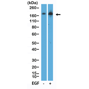
- Scientific DescriptionEGFR (Epidermal growth factor receptor, HER1, ErbB1) belongs to the HER/ERbB family of proteins that includes three other receptor tyrosine kinases, ERbB2, ERbB3, ERbB4. EGFR is a transmembrane receptor and binding of its cognate ligands such as EGF (Epidermal Growth Factor) and TGF alpha (Transforming Growth Factor alpha) to the extracellular domain leads to EGFR dimerization followed by autophosphorylation of the tyrosine residues in the cytoplasmic domain (these include Y992, Y1045, Y1068, Y1148 and Y1173). Phosphorylation of EGF receptor (EGFR) at Tyr845 in the kinase domain is implicated in stabilizing the activation loop, maintaining the active state enzyme, and providing a binding surface for substrate proteins. The SH2 domain of PLCgamma binds at phospho-Tyr992, resulting in activation of PLCgamma-mediated downstream signaling. Phosphorylation of EGFR at Tyr1045 creates a major docking site for the adaptor protein c-Cbl, leading to receptor ubiquitination and degradation following EGFR activation. The GRB2 adaptor protein binds activated EGFR at phospho-Tyr1068. A pair of phosphorylated EGFR residues (Tyr1148 and Tyr1173) provide a docking site for the Shc scaffold protein, with both sites involved in MAP kinase signaling activation. Phosphorylation of EGFR at specific serine and threonine residues attenuates EGFR kinase activity. EGFR carboxy-terminal residues Ser1046 and Ser1047 are phosphorylated by CaM kinase II; mutation of either of these serines results in upregulated EGFR tyrosine autophosphorylation. EGFR activation signals multiple downstream signaling cascades such as the Ras - ERK, PI3K - Akt, Jak - STAT and PKC pathways that help in growth and proliferation of cells. Mutations in the EGFR gene are associated with lung cancer and multiple alternatively spliced transcript variants encode different protein isoforms of EGFR have been found. Increased production or activation of EGFR has been associated with poor prognosis in a variety of tumors. Moreover, EGFR overexpression is observed in tumors of the head and neck, brain, bladder, stomach, breast, lung, endometrium, cervix, vulva, ovary, esophagus, stomach and in squamous cell carcinoma. Deficient signaling of the EGFR and other receptor tyrosine kinases in humans is associated with diseases such as Alzheimers. - Recombinant Antibody. This antibody reacts to human EGF Receptor only when phosphorylated at Y1068. Apllication: WB. Liquid. 50% Glycerol/PBS with 1% BSA and 0.09% sodium azide. EGFR (Epidermal growth factor receptor, HER1, ErbB1) belongs to the HER/ERbB family of proteins that includes three other receptor tyrosine kinases, ERbB2, ERbB3, ERbB4. EGFR is a transmembrane receptor and binding of its cognate ligands such as EGF (Epidermal Growth Factor) and TGF alpha (Transforming Growth Factor alpha) to the extracellular domain leads to EGFR dimerization followed by autophosphorylation of the tyrosine residues in the cytoplasmic domain (these include Y992, Y1045, Y1068, Y1148 and Y1173). Phosphorylation of EGF receptor (EGFR) at Tyr845 in the kinase domain is implicated in stabilizing the activation loop, maintaining the active state enzyme, and providing a binding surface for substrate proteins. The SH2 domain of PLCgamma binds at phospho-Tyr992, resulting in activation of PLCgamma-mediated downstream signaling. Phosphorylation of EGFR at Tyr1045 creates a major docking site for the adaptor protein c-Cbl, leading to receptor ubiquitination and degradation following EGFR activation. The GRB2 adaptor protein binds activated EGFR at phospho-Tyr1068. A pair of phosphorylated EGFR residues (Tyr1148 and Tyr1173) provide a docking site for the Shc scaffold protein, with both sites involved in MAP kinase signaling activation. Phosphorylation of EGFR at specific serine and threonine residues attenuates EGFR kinase activity. EGFR carboxy-terminal residues Ser1046 and Ser1047 are phosphorylated by CaM kinase II; mutation of either of these serines results in upregulated EGFR tyrosine autophosphorylation. EGFR activation signals multiple downstream signaling cascades such as the Ras - ERK, PI3K - Akt, Jak - STAT and PKC pathways that help in growth and proliferation of cells. Mutations in the EGFR gene are associated with lung cancer and multiple alternatively spliced transcript variants encode different protein isoforms of EGFR have been found. Increased production or activation of EGFR has been associated with poor prognosis in a variety of tumors. Moreover, EGFR overexpression is observed in tumors of the head and neck, brain, bladder, stomach, breast, lung, endometrium, cervix, vulva, ovary, esophagus, stomach and in squamous cell carcinoma. Deficient signaling of the EGFR and other receptor tyrosine kinases in humans is associated with diseases such as Alzheimers.
- ReactivityHuman
- Storage Instruction-20°C,2°C to 8°C
- UNSPSC41116161
Datasheet
Related products
Product group Antibodies
ApplicationsImmunoFluorescence, Western Blot, ELISA, ImmunoHistoChemistry
ReactivityHuman, Mouse, Rat
- SizePrice
Product group Antibodies

Anti-Alpha-Tubulin [F2C]Ab00403-1.1
ApplicationsImmunoFluorescence, Western Blot, ELISA
ReactivityHuman
TargetTUBA1A
- SizePrice
Product group Antibodies
Anti-Tubulin alpha Antibody102-25843
ApplicationsImmunoFluorescence, Western Blot, ImmunoHistoChemistry
TargetTUBA1A
- SizePrice

Product group Antibodies
Anti-Tubulin alpha Antibody Picoband(r)A03989-1-CARRIER-FREE
ApplicationsFlow Cytometry, ImmunoFluorescence, Western Blot, ELISA, ImmunoCytoChemistry, ImmunoHistoChemistry
ReactivityHuman, Mouse, Rat
TargetTUBA1A
- SizePrice
Product group Antibodies
Tubulin alpha Polyclonal AntibodyBS-20496R
ApplicationsImmunoFluorescence, Western Blot, ELISA, ImmunoCytoChemistry, ImmunoHistoChemistry, ImmunoHistoChemistry Frozen, ImmunoHistoChemistry Paraffin
ReactivityHuman, Mouse, Rat
TargetTUBA1A
- SizePrice
Product group Antibodies
TUBA1A Monoclonal AntibodyCSB-MA000190
ApplicationsImmunoPrecipitation, Western Blot, ELISA
ReactivityHuman, Mouse, Rat
TargetTUBA1A
- SizePrice
Product group Antibodies
TUBA1A Monoclonal AntibodyCAC12980
ApplicationsFlow Cytometry, ImmunoFluorescence, ImmunoPrecipitation, Western Blot, ELISA, ImmunoHistoChemistry
ReactivityMouse, Rabbit, Rat
- SizePrice
Product group Antibodies

TUBA1A / Tubulin Alpha 1a Antibody (HRP)LS-C377875
ApplicationsELISA, ImmunoHistoChemistry
ReactivityHuman
TargetTUBA1A
- SizePrice

Product group Antibodies
Anti-TUBA1A AntibodyHPA039247
ApplicationsWestern Blot, ImmunoCytoChemistry, ImmunoHistoChemistry
ReactivityHuman, Mouse, Rat
TargetTUBA1A
- SizePrice
Product group Antibodies
alpha Tubulin 1A antibodyGTX109832
ApplicationsImmunoFluorescence, Western Blot, ImmunoCytoChemistry, ImmunoHistoChemistry, ImmunoHistoChemistry Paraffin
ReactivityDrosophila, Human, Mouse, Rat
TargetTUBA1A
- SizePrice