


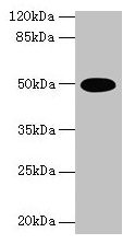
Figure 1. Western blot analysis of TUFM using anti-TUFM antibody (A05606-2). Electrophoresis was performed on a 5-20% SDS-PAGE gel at 70V (Stacking gel) / 90V (Resolving gel) for 2-3 hours. The sample well of each lane was loaded with 30 ug of sample under reducing conditions. Lane 1: human HepG2 whole cell lysates, Lane 2: human Daudi whole cell lysates, Lane 3: human 293T whole cell lysates, Lane 4: human MCF-7 whole cell lysates, Lane 5: rat brain tissue lysates, Lane 6: mouse brain tissue lysates, Lane 7: mouse spleen tissue lysates. After electrophoresis, proteins were transferred to a nitrocellulose membrane at 150 mA for 50-90 minutes. Blocked the membrane with 5% non-fat milk/TBS for 1.5 hour at RT. The membrane was incubated with rabbit anti-TUFM antigen affinity purified polyclonal antibody (Catalog # A05606-2) at 0.25 microg/mL overnight at 4°C, then washed with TBS-0.1%Tween 3 times with 5 minutes each and probed with a goat anti-rabbit IgG-HRP secondary antibody at a dilution of 1:5000 for 1.5 hour at RT. The signal is developed using an Enhanced Chemiluminescent detection (ECL) kit (Catalog # EK1002) with Tanon 5200 system. A specific band was detected for TUFM at approximately 50 kDa. The expected band size for TUFM is at 46 kDa.
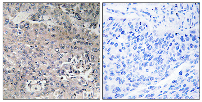
. TUFM was detected in an immunocytochemical section of Hela cells. Enzyme antigen retrieval was performed using IHC enzyme antigen retrieval reagent (AR0022) for 15 mins. The cells were blocked with 10% goat serum. And then incubated with 5 microg/mL rabbit anti-TUFM Antibody (A05606-2) overnight at 4°C. Cy3 Conjugated Goat Anti-Rabbit IgG (BA1032) was used as secondary antibody at 1:500 dilution and incubated for 30 minutes at 37°C. The section was counterstained with DAPI. Visualize using a fluorescence microscope and filter sets appropriate for the label used.")
Figure 1. Western blot analysis of TUFM using anti-TUFM antibody (A05606-2). Electrophoresis was performed on a 5-20% SDS-PAGE gel at 70V (Stacking gel) / 90V (Resolving gel) for 2-3 hours. The sample well of each lane was loaded with 30 ug of sample under reducing conditions. Lane 1: human HepG2 whole cell lysates, Lane 2: human Daudi whole cell lysates, Lane 3: human 293T whole cell lysates, Lane 4: human MCF-7 whole cell lysates, Lane 5: rat brain tissue lysates, Lane 6: mouse brain tissue lysates, Lane 7: mouse spleen tissue lysates. After electrophoresis, proteins were transferred to a nitrocellulose membrane at 150 mA for 50-90 minutes. Blocked the membrane with 5% non-fat milk/TBS for 1.5 hour at RT. The membrane was incubated with rabbit anti-TUFM antigen affinity purified polyclonal antibody (Catalog # A05606-2) at 0.25 microg/mL overnight at 4°C, then washed with TBS-0.1%Tween 3 times with 5 minutes each and probed with a goat anti-rabbit IgG-HRP secondary antibody at a dilution of 1:5000 for 1.5 hour at RT. The signal is developed using an Enhanced Chemiluminescent detection (ECL) kit (Catalog # EK1002) with Tanon 5200 system. A specific band was detected for TUFM at approximately 50 kDa. The expected band size for TUFM is at 46 kDa.
Anti-TUFM Antibody Picoband(r)
A05606-2-HRP
ApplicationsImmunoFluorescence, Western Blot, ELISA, ImmunoCytoChemistry
Product group Antibodies
ReactivityHuman, Mouse, Rat
TargetTUFM
Overview
- SupplierBoster Bio
- Product NameAnti-TUFM Antibody Picoband(r)
- Delivery Days Customer9
- ApplicationsImmunoFluorescence, Western Blot, ELISA, ImmunoCytoChemistry
- CertificationResearch Use Only
- ClonalityPolyclonal
- Concentration500 ug/ml
- ConjugateHRP
- Gene ID7284
- Target nameTUFM
- Target descriptionTu translation elongation factor, mitochondrial
- Target synonymsCOXPD4, EF-TuMT, EFTU, P43, elongation factor Tu, mitochondrial, EF-Tu, epididymis secretory sperm binding protein
- HostRabbit
- IsotypeIgG
- Protein IDP49411
- Protein NameElongation factor Tu, mitochondrial
- Scientific DescriptionBoster Bio Anti-TUFM Antibody Picoband® catalog # A05606-2. Tested in ELISA, IF, ICC, WB applications. This antibody reacts with Human, Mouse, Rat. The brand Picoband indicates this is a premium antibody that guarantees superior quality, high affinity, and strong signals with minimal background in Western blot applications. Only our best-performing antibodies are designated as Picoband, ensuring unmatched performance.
- ReactivityHuman, Mouse, Rat
- Storage Instruction-20°C,2°C to 8°C
- UNSPSC12352203
Related products
Product group Antibodies
TUFM Polyclonal AntibodyCAC13937
ApplicationsWestern Blot, ELISA
TargetTUFM
- SizePrice

Product group Antibodies
Anti-TUFM AntibodyAMAB90964
ApplicationsWestern Blot, ImmunoCytoChemistry, ImmunoHistoChemistry
ReactivityHuman
TargetTUFM
- SizePrice
Product group Antibodies
Anti-TUFM Antibody144-06423
ApplicationsImmunoFluorescence, ImmunoPrecipitation, Western Blot, ImmunoHistoChemistry
ReactivityHuman, Mouse, Rat
TargetTUFM
- SizePrice
Product group Antibodies
Anti-TUFM AntibodyA97759
ApplicationsELISA, ImmunoHistoChemistry
ReactivityHuman, Mouse, Rat
- SizePrice
Product group Antibodies
References
TUFM antibodyGTX101763
ApplicationsImmunoFluorescence, ImmunoPrecipitation, Western Blot, ImmunoCytoChemistry, ImmunoHistoChemistry, ImmunoHistoChemistry Paraffin
ReactivityHuman, Mouse
TargetTUFM
- SizePrice
Product group Antibodies

EFTU / TUFM AntibodyLS-C771507
ApplicationsWestern Blot
ReactivityHuman, Rat
TargetTUFM
- SizePrice
Product group Antibodies
Anti-TUFM Antibody Picoband(r)A05606-2-CARRIER-FREE
ApplicationsImmunoFluorescence, Western Blot, ELISA, ImmunoCytoChemistry
ReactivityHuman, Mouse, Rat
TargetTUFM
- SizePrice
Product group Antibodies
TUFM AntibodyCSB-PA02199A0RB
ApplicationsWestern Blot, ELISA
ReactivityHuman
TargetTUFM
- SizePrice