


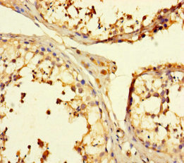
Immunohistochemical staining of human skin shows cytoplasmic positivity in squamous epithelial cells.
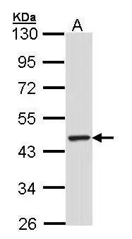
 and TULP3 over-expression lysate (Co-expressed with a C-terminal myc-DDK tag (~3.1 kDa) in mammalian HEK293T cells, LY418767).")
Immunohistochemical staining of human skin shows cytoplasmic positivity in squamous epithelial cells.
Anti-TULP3 Antibody
HPA015285
ApplicationsWestern Blot, ImmunoHistoChemistry
Product group Antibodies
ReactivityHuman
TargetTULP3
Overview
- SupplierAtlas Antibodies
- Product NameAnti-TULP3 Antibody
- Delivery Days Customer4
- ApplicationsWestern Blot, ImmunoHistoChemistry
- CertificationResearch Use Only
- ClonalityPolyclonal
- ConjugateUnconjugated
- Gene ID7289
- Target nameTULP3
- Target descriptionTUB like protein 3
- Target synonymsHRCDF, TUBL3, tubby-related protein 3, tubby like protein 3
- HostRabbit
- IsotypeIgG
- Protein IDO75386
- Protein NameTubby-related protein 3
- Scientific DescriptionRecombinant Protein Epitope Signature Tag (PrEST) antigen sequence
- ReactivityHuman
- Storage Instruction-20°C,2°C to 8°C
- UNSPSC41116161
Datasheet
MSDS
Related products
Product group Antibodies
TULP3 AntibodyCSB-PA025348LA01HU
ApplicationsELISA, ImmunoHistoChemistry
ReactivityHuman
TargetTULP3
- SizePrice

Product group Antibodies
Anti-TULP3 Antibody Picoband(r)A09384-2-CARRIER-FREE
ApplicationsWestern Blot, ELISA
ReactivityHuman
TargetTULP3
- SizePrice

Product group Antibodies
Anti-TULP3 AntibodyHPA018496
ApplicationsImmunoCytoChemistry
ReactivityHuman
TargetTULP3
- SizePrice
Product group Antibodies
Anti-TULP3 AntibodyHPA018496
ApplicationsImmunoCytoChemistry
ReactivityHuman
TargetTULP3
- SizePrice
Product group Antibodies

TULP3 Antibody (HRP)LS-C394744
ApplicationsELISA
ReactivityHuman
TargetTULP3
- SizePrice
Product group Antibodies
TULP3 antibody [N2C3]GTX104995
ApplicationsImmunoFluorescence, Western Blot, ImmunoCytoChemistry
ReactivityHuman
TargetTULP3
- SizePrice