


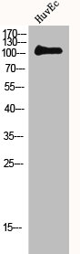
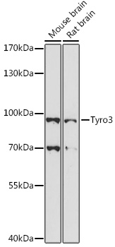
Figure 1. Western blot analysis of TYRO3 using anti-TYRO3 antibody (M00913-5). Electrophoresis was performed on a 5-20% SDS-PAGE gel at 70V (Stacking gel) / 90V (Resolving gel) for 2-3 hours. The sample well of each lane was loaded with 30 ug of sample under reducing conditions. Lane 1: human MCF-7 whole cell lysates, Lane 2: human U251 whole cell lysates, Lane 3: human HepG2 whole cell lysates, Lane 4: rat brain tissue lysates, Lane 5: mouse brain tissue lysates, Lane 6: mouse lung tissue lysates. After electrophoresis, proteins were transferred to a nitrocellulose membrane at 150 mA for 50-90 minutes. Blocked the membrane with 5% non-fat milk/TBS for 1.5 hour at RT. The membrane was incubated with rabbit anti-TYRO3 antigen affinity purified monoclonal antibody (Catalog # M00913-5) at 1:500 overnight at 4°C, then washed with TBS-0.1%Tween 3 times with 5 minutes each and probed with a goat anti-rabbit IgG-HRP secondary antibody at a dilution of 1:5000 for 1.5 hour at RT. The signal is developed using an Enhanced Chemiluminescent detection (ECL) kit (Catalog # EK1002) with Tanon 5200 system. A specific band was detected for TYRO3 at approximately 120 kDa. The expected band size for TYRO3 is at 97 kDa.
Figure 1. Western blot analysis of TYRO3 using anti-TYRO3 antibody (M00913-5). Electrophoresis was performed on a 5-20% SDS-PAGE gel at 70V (Stacking gel) / 90V (Resolving gel) for 2-3 hours. The sample well of each lane was loaded with 30 ug of sample under reducing conditions. Lane 1: human MCF-7 whole cell lysates, Lane 2: human U251 whole cell lysates, Lane 3: human HepG2 whole cell lysates, Lane 4: rat brain tissue lysates, Lane 5: mouse brain tissue lysates, Lane 6: mouse lung tissue lysates. After electrophoresis, proteins were transferred to a nitrocellulose membrane at 150 mA for 50-90 minutes. Blocked the membrane with 5% non-fat milk/TBS for 1.5 hour at RT. The membrane was incubated with rabbit anti-TYRO3 antigen affinity purified monoclonal antibody (Catalog # M00913-5) at 1:500 overnight at 4°C, then washed with TBS-0.1%Tween 3 times with 5 minutes each and probed with a goat anti-rabbit IgG-HRP secondary antibody at a dilution of 1:5000 for 1.5 hour at RT. The signal is developed using an Enhanced Chemiluminescent detection (ECL) kit (Catalog # EK1002) with Tanon 5200 system. A specific band was detected for TYRO3 at approximately 120 kDa. The expected band size for TYRO3 is at 97 kDa.
Anti-TYRO3 Rabbit Monoclonal Antibody
M00913-5
ApplicationsImmunoFluorescence, ImmunoPrecipitation, Western Blot, ImmunoCytoChemistry, ImmunoHistoChemistry
Product group Antibodies
ReactivityHuman, Mouse, Rat
TargetTYRO3
Overview
- SupplierBoster Bio
- Product NameAnti-TYRO3 Rabbit Monoclonal Antibody
- Delivery Days Customer9
- ApplicationsImmunoFluorescence, ImmunoPrecipitation, Western Blot, ImmunoCytoChemistry, ImmunoHistoChemistry
- CertificationResearch Use Only
- ClonalityMonoclonal
- Clone ID22T29
- Gene ID7301
- Target nameTYRO3
- Target descriptionTYRO3 protein tyrosine kinase
- Target synonymsBYK, Dtk, Etk-2, RSE, Rek, Sky, Tif, tyrosine-protein kinase receptor TYRO3, tyrosine-protein kinase DTK, tyrosine-protein kinase RSE, tyrosine-protein kinase SKY, tyrosine-protein kinase TIF, tyrosine-protein kinase byk
- HostRabbit
- IsotypeIgG
- Protein IDQ06418
- Protein NameTyrosine-protein kinase receptor TYRO3
- Scientific DescriptionBoster Bio Anti-TYRO3 Rabbit Monoclonal Antibody catalog # M00913-5. Tested in WB, IHC, ICC/IF, IP applications. This antibody reacts with Human, Mouse, Rat.
- ReactivityHuman, Mouse, Rat
- Storage Instruction-20°C
- UNSPSC12352203
Related products
Product group Antibodies
ReactivityHuman
TargetTYRO3
- SizePrice
Product group Antibodies
MERTK/TYRO3 AntibodyCSB-PA003230
ApplicationsWestern Blot, ELISA
ReactivityHuman, Mouse, Rat
TargetTYRO3
- SizePrice
Product group Antibodies
Anti-TYRO3 Antibody144-63157
ApplicationsWestern Blot
ReactivityHuman, Mouse, Rat
TargetTYRO3
- SizePrice
Product group Antibodies
Anti-TYRO3 AntibodyA91424
ApplicationsWestern Blot
ReactivityMouse, Rat
- SizePrice
Product group Antibodies

TYRO3 AntibodyLS-C747394
ApplicationsWestern Blot
ReactivityHuman, Mouse, Rat
TargetTYRO3
- SizePrice
Product group Antibodies
TYRO3 Recombinant Antibody, AbBy Fluor-405 ConjugatedBSM-62103R-BF405
ApplicationsImmunoFluorescence, Western Blot
ReactivityHuman, Mouse, Rat
TargetTYRO3
- SizePrice