


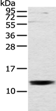
Figure 1. Western blot analysis of UQCRH using anti-UQCRH antibody (M09436). Electrophoresis was performed on a 5-20% SDS-PAGE gel at 70V (Stacking gel) / 90V (Resolving gel) for 2-3 hours. The sample well of each lane was loaded with 30 ug of sample under reducing conditions. Lane 1: human K562 whole cell lysates, Lane 2: human THP-1 whole cell lysates, Lane 3: rat brain tissue lysates, Lane 4: rat stomach tissue lysates, Lane 5: mouse brain tissue lysates, Lane 6: mouse stomach tissue lysates. After electrophoresis, proteins were transferred to a nitrocellulose membrane at 150 mA for 50-90 minutes. Blocked the membrane with 5% non-fat milk/TBS for 1.5 hour at RT. The membrane was incubated with rabbit anti-UQCRH antigen affinity purified monoclonal antibody (Catalog # M09436) at 1:500 overnight at 4°C, then washed with TBS-0.1%Tween 3 times with 5 minutes each and probed with a goat anti-rabbit IgG-HRP secondary antibody at a dilution of 1:500 for 1.5 hour at RT. The signal is developed using an Enhanced Chemiluminescent detection (ECL) kit (Catalog # EK1002) with Tanon 5200 system. A specific band was detected for UQCRH at approximately 13 kDa. The expected band size for UQCRH is at 11 kDa.
Figure 1. Western blot analysis of UQCRH using anti-UQCRH antibody (M09436). Electrophoresis was performed on a 5-20% SDS-PAGE gel at 70V (Stacking gel) / 90V (Resolving gel) for 2-3 hours. The sample well of each lane was loaded with 30 ug of sample under reducing conditions. Lane 1: human K562 whole cell lysates, Lane 2: human THP-1 whole cell lysates, Lane 3: rat brain tissue lysates, Lane 4: rat stomach tissue lysates, Lane 5: mouse brain tissue lysates, Lane 6: mouse stomach tissue lysates. After electrophoresis, proteins were transferred to a nitrocellulose membrane at 150 mA for 50-90 minutes. Blocked the membrane with 5% non-fat milk/TBS for 1.5 hour at RT. The membrane was incubated with rabbit anti-UQCRH antigen affinity purified monoclonal antibody (Catalog # M09436) at 1:500 overnight at 4°C, then washed with TBS-0.1%Tween 3 times with 5 minutes each and probed with a goat anti-rabbit IgG-HRP secondary antibody at a dilution of 1:500 for 1.5 hour at RT. The signal is developed using an Enhanced Chemiluminescent detection (ECL) kit (Catalog # EK1002) with Tanon 5200 system. A specific band was detected for UQCRH at approximately 13 kDa. The expected band size for UQCRH is at 11 kDa.
Anti-UQCRH Rabbit Monoclonal Antibody
M09436
ApplicationsWestern Blot, ImmunoHistoChemistry
Product group Antibodies
ReactivityHuman, Mouse, Rat
TargetUQCRH
Overview
- SupplierBoster Bio
- Product NameAnti-UQCRH Rabbit Monoclonal Antibody
- Delivery Days Customer9
- ApplicationsWestern Blot, ImmunoHistoChemistry
- CertificationResearch Use Only
- ClonalityMonoclonal
- Clone ID27U84
- Gene ID7388
- Target nameUQCRH
- Target descriptionubiquinol-cytochrome c reductase hinge protein
- Target synonymsMC3DN11, QCR6, UQCR8, cytochrome b-c1 complex subunit 6, mitochondrial, complex III subunit 6, cytochrome c1 non-heme 11 kDa protein, mitochondrial hinge protein, ubiquinol-cytochrome c reductase complex 11 kDa protein, ubiquinol-cytochrome c reductase, complex III subunit VIII
- HostRabbit
- IsotypeIgG
- Protein IDP07919
- Protein NameCytochrome b-c1 complex subunit 6, mitochondrial
- Scientific DescriptionBoster Bio Anti-UQCRH Rabbit Monoclonal Antibody catalog # M09436. Tested in WB, IHC applications. This antibody reacts with Human, Mouse, Rat.
- ReactivityHuman, Mouse, Rat
- Storage Instruction-20°C
- UNSPSC12352203
Related products
Product group Antibodies
UQCRH AntibodyCSB-PA025671GA01HU
ApplicationsELISA
ReactivityHuman, Mouse, Rat
TargetUQCRH
- SizePrice
Product group Antibodies
Anti-UQCRH AntibodyA43479
ApplicationsWestern Blot
ReactivityHuman, Mouse
- SizePrice

Product group Antibodies
Anti-UQCRH AntibodyHPA042574
ApplicationsImmunoHistoChemistry
ReactivityHuman
TargetUQCRH
- SizePrice
Product group Antibodies

UQCRH AntibodyLS-C661462
ApplicationsELISA, ImmunoHistoChemistry
ReactivityHuman
TargetUQCRH
- SizePrice
Product group Antibodies
UQCRH Recombinant AntibodyBSM-62544R
ApplicationsImmunoFluorescence, Western Blot, ImmunoHistoChemistry, ImmunoHistoChemistry Frozen, ImmunoHistoChemistry Paraffin
ReactivityHuman, Mouse, Rat
TargetUQCRH
- SizePrice