


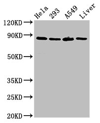
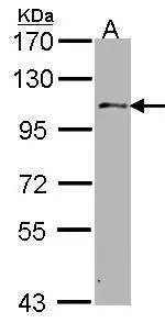
Western blot analysis of USP10 expression in A375 cell lysate.
Western blot analysis of USP10 expression in A375 cell lysate.
Anti-USP10 Monoclonal Antibody
M03786
ApplicationsImmunoFluorescence, ImmunoPrecipitation, Western Blot, ImmunoCytoChemistry, ImmunoHistoChemistry
Product group Antibodies
ReactivityHuman
TargetUSP10
Overview
- SupplierBoster Bio
- Product NameAnti-USP10 Monoclonal Antibody
- Delivery Days Customer9
- ApplicationsImmunoFluorescence, ImmunoPrecipitation, Western Blot, ImmunoCytoChemistry, ImmunoHistoChemistry
- CertificationResearch Use Only
- ClonalityMonoclonal
- Clone IDADCB-21
- Gene ID9100
- Target nameUSP10
- Target descriptionubiquitin specific peptidase 10
- Target synonymsUBPO, ubiquitin carboxyl-terminal hydrolase 10, deubiquitinating enzyme 10, ubiquitin specific protease 10, ubiquitin thioesterase 10, ubiquitin thiolesterase 10, ubiquitin-specific-processing protease 10
- HostRabbit
- IsotypeIgG
- Protein IDQ14694
- Protein NameUbiquitin carboxyl-terminal hydrolase 10
- Scientific DescriptionBoster Bio Anti-USP10 Monoclonal Antibody catalog # M03786. Tested in WB, IHC, ICC/IF, IP applications. This antibody reacts with Human.
- ReactivityHuman
- Storage Instruction-20°C
- UNSPSC12352203
Datasheet
MSDS
Related products

Product group Antibodies
Anti-USP10 Antibody Picoband(r)A03786-3-CARRIER-FREE
ApplicationsWestern Blot, ELISA
ReactivityHuman, Mouse, Rat
TargetUSP10
- SizePrice
Product group Antibodies
Anti-USP10 Antibody144-07505
ApplicationsImmunoFluorescence, Western Blot
ReactivityHuman, Mouse
TargetUSP10
- SizePrice
Product group Antibodies
Anti-USP10 AntibodyA32124
ApplicationsWestern Blot, ImmunoHistoChemistry
ReactivityHuman, Mouse, Rat
- SizePrice
Product group Antibodies
USP10 Recombinant Antibody, AbBy Fluor-350 ConjugatedBSM-61654R-BF350
ApplicationsImmunoFluorescence, Western Blot
ReactivityHuman
TargetUSP10
- SizePrice
Product group Antibodies
USP10 AntibodyCSB-PA613516LA01HU
ApplicationsImmunoFluorescence, ImmunoPrecipitation, Western Blot, ELISA, ImmunoHistoChemistry
ReactivityHuman, Rat
TargetUSP10
- SizePrice
Product group Antibodies
Usp10 Polyclonal AntibodyCAC11210
ApplicationsImmunoFluorescence, ImmunoPrecipitation, Western Blot, ELISA, ImmunoHistoChemistry
ReactivityRat
TargetUSP10
- SizePrice

Product group Antibodies
Anti-USP10 AntibodyHPA006731
ApplicationsWestern Blot, ImmunoCytoChemistry, ImmunoHistoChemistry
ReactivityHuman
TargetUSP10
- SizePrice
Product group Antibodies
USP10 antibody [C2C3], C-termGTX106092
ApplicationsWestern Blot
ReactivityHuman
TargetUSP10
- SizePrice
Product group Antibodies

USP10 AntibodyLS-C748438
ApplicationsImmunoFluorescence, ImmunoHistoChemistry
ReactivityHuman, Mouse
TargetUSP10
- SizePrice