


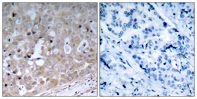
Figure 2. IHC analysis of VEGF Receptor 2 using anti-VEGF Receptor 2 antibody (A00901-2). VEGF Receptor 2 was detected in paraffin-embedded section of human intestinal cancer tissue. Heat mediated antigen retrieval was performed in citrate buffer (pH6, epitope retrieval solution) for 20 mins. The tissue section was blocked with 10% goat serum. The tissue section was then incubated with 1ug/ml rabbit anti-VEGF Receptor 2 Antibody (A00901-2) overnight at 4 Biotinylated goat anti-rabbit IgG was used as secondary antibody and incubated for 30 minutes at 37 The tissue section was developed using Strepavidin-Biotin-Complex (SABC)(Catalog # SA1022) with DAB as the chromogen.
. VEGF Receptor 2 was detected in paraffin-embedded section of human lung cancer tissue. Heat mediated antigen retrieval was performed in citrate buffer (pH6, epitope retrieval solution) for 20 mins. The tissue section was blocked with 10% goat serum. The tissue section was then incubated with 1ug/ml rabbit anti-VEGF Receptor 2 Antibody (A00901-2) overnight at 4 Biotinylated goat anti-rabbit IgG was used as secondary antibody and incubated for 30 minutes at 37 The tissue section was developed using Strepavidin-Biotin-Complex (SABC)(Catalog # SA1022) with DAB as the chromogen.")
. VEGF Receptor 2 was detected in paraffin-embedded section of human placenta tissue. Heat mediated antigen retrieval was performed in citrate buffer (pH6, epitope retrieval solution) for 20 mins. The tissue section was blocked with 10% goat serum. The tissue section was then incubated with 1ug/ml rabbit anti-VEGF Receptor 2 Antibody (A00901-2) overnight at 4 Biotinylated goat anti-rabbit IgG was used as secondary antibody and incubated for 30 minutes at 37 The tissue section was developed using Strepavidin-Biotin-Complex (SABC)(Catalog # SA1022) with DAB as the chromogen.")
. VEGF Receptor 2 was detected in paraffin-embedded section of human mammary cancer tissue. Heat mediated antigen retrieval was performed in citrate buffer (pH6, epitope retrieval solution) for 20 mins. The tissue section was blocked with 10% goat serum. The tissue section was then incubated with 1ug/ml rabbit anti-VEGF Receptor 2 Antibody (A00901-2) overnight at 4 Biotinylated goat anti-rabbit IgG was used as secondary antibody and incubated for 30 minutes at 37 The tissue section was developed using Strepavidin-Biotin-Complex (SABC)(Catalog # SA1022) with DAB as the chromogen.")
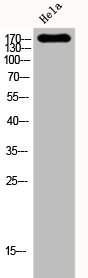
. Electrophoresis was performed on a 5-20% SDS-PAGE gel at 70V (Stacking gel) / 90V (Resolving gel) for 2-3 hours. The sample well of each lane was loaded with 30 ug of sample under reducing conditions. Lane 1: human placenta tissue lysates, Lane 2: human PC-3 whole cell lysates. After electrophoresis, proteins were transferred to a nitrocellulose membrane at 150 mA for 50-90 minutes. Blocked the membrane with 5% non-fat milk/TBS for 1.5 hour at RT. The membrane was incubated with rabbit anti-VEGF Receptor 2 antigen affinity purified polyclonal antibody (Catalog # A00901-2) at 0.5 microg/mL overnight at 4°C, then washed with TBS-0.1%Tween 3 times with 5 minutes each and probed with a goat anti-rabbit IgG-HRP secondary antibody at a dilution of 1:5000 for 1.5 hour at RT. The signal is developed using an Enhanced Chemiluminescent detection (ECL) kit (Catalog # EK1002) with Tanon 5200 system. A specific band was detected for VEGF Receptor 2 at approximately 180-250 kDa. The expected band size for VEGF Receptor 2 is at 156 kDa.")
Figure 2. IHC analysis of VEGF Receptor 2 using anti-VEGF Receptor 2 antibody (A00901-2). VEGF Receptor 2 was detected in paraffin-embedded section of human intestinal cancer tissue. Heat mediated antigen retrieval was performed in citrate buffer (pH6, epitope retrieval solution) for 20 mins. The tissue section was blocked with 10% goat serum. The tissue section was then incubated with 1ug/ml rabbit anti-VEGF Receptor 2 Antibody (A00901-2) overnight at 4 Biotinylated goat anti-rabbit IgG was used as secondary antibody and incubated for 30 minutes at 37 The tissue section was developed using Strepavidin-Biotin-Complex (SABC)(Catalog # SA1022) with DAB as the chromogen.
Anti-VEGF Receptor 2/KDR Antibody Picoband(r)
A00901-2-CARRIER-FREE
ApplicationsFlow Cytometry, Western Blot, ImmunoCytoChemistry, ImmunoHistoChemistry
Product group Antibodies
ReactivityHuman
TargetKDR
Overview
- SupplierBoster Bio
- Product NameAnti-VEGF Receptor 2/KDR Antibody Picoband(r)
- Delivery Days Customer9
- ApplicationsFlow Cytometry, Western Blot, ImmunoCytoChemistry, ImmunoHistoChemistry
- CertificationResearch Use Only
- ClonalityPolyclonal
- Concentration500 ug/ml
- Gene ID3791
- Target nameKDR
- Target descriptionkinase insert domain receptor
- Target synonymsCD309, FLK1, VEGFR, VEGFR2, vascular endothelial growth factor receptor 2, fetal liver kinase-1, kinase insert domain receptor (a type III receptor tyrosine kinase), protein-tyrosine kinase receptor Flk-1, soluble VEGFR2, tyrosine kinase growth factor receptor
- HostRabbit
- IsotypeIgG
- Protein IDP35968
- Protein NameVascular endothelial growth factor receptor 2
- Scientific DescriptionBoster Bio Anti-VEGF Receptor 2/KDR Antibody Picoband® catalog # A00901-2. Tested in Flow Cytometry, IHC, ICC, WB applications. This antibody reacts with Human. The brand Picoband indicates this is a premium antibody that guarantees superior quality, high affinity, and strong signals with minimal background in Western blot applications. Only our best-performing antibodies are designated as Picoband, ensuring unmatched performance.
- ReactivityHuman
- Storage Instruction-20°C,2°C to 8°C
- UNSPSC12352203
Related products
Product group Antibodies
Anti-VEGFR2 AntibodyA95579
ApplicationsWestern Blot, ELISA, ImmunoHistoChemistry
ReactivityHuman, Mouse, Rat
- SizePrice
Product group Antibodies
anti-VEGFR-2/KDR (human), mAb (3(4H3))AG-20T-0109
ApplicationsFlow Cytometry, Western Blot
ReactivityHuman
TargetKDR
- SizePrice
Product group Antibodies

Anti-VEGFR2 [IMC-1121B (Ramucirumab)]Ab03058-10.0
ApplicationsFlow Cytometry, ImmunoHistoChemistry, Other Application
ReactivityHuman
TargetKDR
- SizePrice
Product group Antibodies
Anti-KDR Antibody144-11127
ApplicationsWestern Blot, ImmunoHistoChemistry
ReactivityHuman, Mouse, Rat
TargetKDR
- SizePrice
Product group Antibodies
References
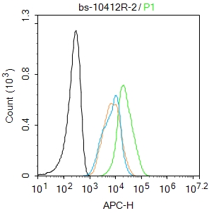
VEGFR2 Polyclonal AntibodyBS-10412R
ApplicationsFlow Cytometry, ImmunoFluorescence, ELISA, ImmunoHistoChemistry, ImmunoHistoChemistry Frozen, ImmunoHistoChemistry Paraffin
TargetKDR
- SizePrice
Product group Antibodies
KDR AntibodyCSB-PA002542
ApplicationsImmunoFluorescence, Western Blot, ELISA, ImmunoHistoChemistry
ReactivityHuman, Mouse
TargetKDR
- SizePrice
Product group Antibodies
ApplicationsImmunoPrecipitation, Western Blot, ImmunoCytoChemistry, ImmunoHistoChemistry
ReactivityMouse, Rat
TargetKDR
- SizePrice
Product group Antibodies

KDR / VEGFR2 / FLK1 Antibody (Internal)LS-C368404
ApplicationsImmunoFluorescence, Western Blot, ImmunoCytoChemistry, ImmunoHistoChemistry, ImmunoHistoChemistry Paraffin
ReactivityHuman
TargetKDR
- SizePrice

Product group Antibodies
Anti-KDR AntibodyHPA073577
ApplicationsImmunoCytoChemistry
ReactivityHuman
TargetKDR
- SizePrice