


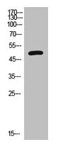
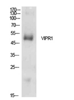
Figure 1. Western blot analysis of VIP Receptor 1 using anti-VIP Receptor 1 antibody (A04345). Electrophoresis was performed on a 5-20% SDS-PAGE gel at 70V (Stacking gel) / 90V (Resolving gel) for 2-3 hours. The sample well of each lane was loaded with 50ug of sample under reducing conditions. Lane 1: human A431 cell lysates, Lane 2: human PANC-1 cell lysates. After Electrophoresis, proteins were transferred to a Nitrocellulose membrane at 150mA for 50-90 minutes. Blocked the membrane with 5% Non-fat Milk/ TBS for 1.5 hour at RT. The membrane was incubated with rabbit anti-VIP Receptor 1 antigen affinity purified polyclonal antibody (Catalog # A04345) at 0.5 microg/mL overnight at 4°C, then washed with TBS-0.1%Tween 3 times with 5 minutes each and probed with a goat anti-rabbit IgG-HRP secondary antibody at a dilution of 1:10000 for 1.5 hour at RT. The signal is developed using an Enhanced Chemiluminescent detection (ECL) kit (Catalog # EK1002) with Tanon 5200 system. A specific band was detected for VIP Receptor 1 at approximately 52KD. The expected band size for VIP Receptor 1 is at 52KD.
Figure 1. Western blot analysis of VIP Receptor 1 using anti-VIP Receptor 1 antibody (A04345). Electrophoresis was performed on a 5-20% SDS-PAGE gel at 70V (Stacking gel) / 90V (Resolving gel) for 2-3 hours. The sample well of each lane was loaded with 50ug of sample under reducing conditions. Lane 1: human A431 cell lysates, Lane 2: human PANC-1 cell lysates. After Electrophoresis, proteins were transferred to a Nitrocellulose membrane at 150mA for 50-90 minutes. Blocked the membrane with 5% Non-fat Milk/ TBS for 1.5 hour at RT. The membrane was incubated with rabbit anti-VIP Receptor 1 antigen affinity purified polyclonal antibody (Catalog # A04345) at 0.5 microg/mL overnight at 4°C, then washed with TBS-0.1%Tween 3 times with 5 minutes each and probed with a goat anti-rabbit IgG-HRP secondary antibody at a dilution of 1:10000 for 1.5 hour at RT. The signal is developed using an Enhanced Chemiluminescent detection (ECL) kit (Catalog # EK1002) with Tanon 5200 system. A specific band was detected for VIP Receptor 1 at approximately 52KD. The expected band size for VIP Receptor 1 is at 52KD.
Anti-VIP Receptor 1/VIPR1 Antibody Picoband(r)
A04345-CARRIER-FREE
ApplicationsWestern Blot, ImmunoHistoChemistry
Product group Antibodies
ReactivityHuman, Mouse, Rat
TargetVIPR1
Overview
- SupplierBoster Bio
- Product NameAnti-VIP Receptor 1/VIPR1 Antibody Picoband(r)
- Delivery Days Customer9
- ApplicationsWestern Blot, ImmunoHistoChemistry
- CertificationResearch Use Only
- ClonalityPolyclonal
- Concentration500 ug/ml
- Gene ID7433
- Target nameVIPR1
- Target descriptionvasoactive intestinal peptide receptor 1
- Target synonymsHVR1, II, PACAP-R-2, PACAP-R2, RDC1, V1RG, VAPC1, VIP-R-1, VIPR, VIRG, VPAC1, VPAC1R, VPCAP1R, vasoactive intestinal polypeptide receptor 1, PACAP type II receptor, VIP and PACAP receptor 1, VIP receptor, type I, VPAC1 receptor, pituitary adenylate cyclase activating polypeptide receptor, type II, type 1 vasoactive intestinal peptide receptor
- HostRabbit
- IsotypeIgG
- Protein IDP32241
- Protein NameVasoactive intestinal polypeptide receptor 1
- Scientific DescriptionBoster Bio Anti-VIP Receptor 1/VIPR1 Antibody Picoband® catalog # A04345. Tested in IHC, WB applications. This antibody reacts with Human, Mouse, Rat. The brand Picoband indicates this is a premium antibody that guarantees superior quality, high affinity, and strong signals with minimal background in Western blot applications. Only our best-performing antibodies are designated as Picoband, ensuring unmatched performance.
- ReactivityHuman, Mouse, Rat
- Storage Instruction-20°C,2°C to 8°C
- UNSPSC12352203
Related products
Product group Antibodies
VIPR1 AntibodyCSB-PA006153
ApplicationsWestern Blot, ELISA
ReactivityHuman
TargetVIPR1
- SizePrice
Product group Antibodies
Anti-VIPR1 AntibodyA100723
ApplicationsWestern Blot, ELISA
ReactivityHuman
- SizePrice
Product group Antibodies
Anti-VIPR1 Antibody144-61921
ApplicationsWestern Blot, ImmunoHistoChemistry
ReactivityHuman, Rat
TargetVIPR1
- SizePrice
Product group Antibodies

VIPR1 AntibodyLS-C497226
ApplicationsWestern Blot
ReactivityHuman, Rat
TargetVIPR1
- SizePrice
Product group Antibodies
References
ApplicationsImmunoFluorescence, Western Blot, ELISA, ImmunoCytoChemistry, ImmunoHistoChemistry, ImmunoHistoChemistry Frozen, ImmunoHistoChemistry Paraffin
ReactivityBovine, Human, Mouse, Porcine, Rabbit, Rat, Sheep
TargetVIPR1
- SizePrice