

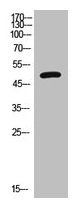
Western Blot analysis of HepG2 cells using VPAC1 Polyclonal Antibody.
Western Blot analysis of HepG2 cells using VPAC1 Polyclonal Antibody.
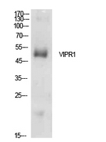
VIPR1 Antibody
CSB-PA006153
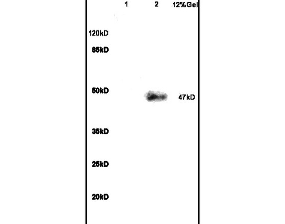
ApplicationsWestern Blot, ELISA
Product group Antibodies
ReactivityHuman
TargetVIPR1
Overview
- SupplierCusabio
- Product NameVIPR1 Antibody
- Delivery Days Customer20
- ApplicationsWestern Blot, ELISA
- CertificationResearch Use Only
- ClonalityPolyclonal
- ConjugateUnconjugated
- Gene ID7433
- Target nameVIPR1
- Target descriptionvasoactive intestinal peptide receptor 1
- Target synonymsHVR1, II, PACAP-R-2, PACAP-R2, RDC1, V1RG, VAPC1, VIP-R-1, VIPR, VIRG, VPAC1, VPAC1R, VPCAP1R, vasoactive intestinal polypeptide receptor 1, PACAP type II receptor, VIP and PACAP receptor 1, VIP receptor, type I, VPAC1 receptor, pituitary adenylate cyclase activating polypeptide receptor, type II, type 1 vasoactive intestinal peptide receptor
- HostRabbit
- IsotypeIgG
- Protein IDP32241
- Protein NameVasoactive intestinal polypeptide receptor 1
- ReactivityHuman
- Storage Instruction-20°C or -80°C
- UNSPSC41116161
Related products
Product group Antibodies
Anti-VIPR1 AntibodyA100723
ApplicationsWestern Blot, ELISA
ReactivityHuman
- SizePrice
Product group Antibodies
Anti-VIPR1 Antibody144-61921
ApplicationsWestern Blot, ImmunoHistoChemistry
ReactivityHuman, Rat
TargetVIPR1
- SizePrice

Product group Antibodies
Anti-VIP Receptor 1/VIPR1 Antibody Picoband(r)A04345-CARRIER-FREE
ApplicationsWestern Blot, ImmunoHistoChemistry
ReactivityHuman, Mouse, Rat
TargetVIPR1
- SizePrice
Product group Antibodies

VIPR1 AntibodyLS-C497226
ApplicationsWestern Blot
ReactivityHuman, Rat
TargetVIPR1
- SizePrice
Product group Antibodies
References
ApplicationsImmunoFluorescence, Western Blot, ELISA, ImmunoCytoChemistry, ImmunoHistoChemistry, ImmunoHistoChemistry Frozen, ImmunoHistoChemistry Paraffin
ReactivityBovine, Human, Mouse, Porcine, Rabbit, Rat, Sheep
TargetVIPR1
- SizePrice