


Immunohistochemical staining of human colon shows strong nuclear positivity in glandular cells.
![Lane 1: Marker [kDa] 230, 130, 95, 72, 56, 36, 28, 17, 11. Lane 2: Human cell line RT-4. Lane 3: Human cell line U-251MG sp. Lane 4: Human plasma (IgG/HSA depleted). Lane 5: Human liver tissue. Lane 6: Human tonsil tissue](https://atlasantibodies.s3.amazonaws.com/images/wb/hpa026897-wb-1.jpg "Lane 1: Marker [kDa] 230, 130, 95, 72, 56, 36, 28, 17, 11. Lane 2: Human cell line RT-4. Lane 3: Human cell line U-251MG sp. Lane 4: Human plasma (IgG/HSA depleted). Lane 5: Human liver tissue. Lane 6: Human tonsil tissue")
Immunohistochemical staining of human colon shows strong nuclear positivity in glandular cells.
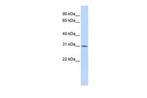
Anti-WDR33 Antibody
HPA026897
ApplicationsWestern Blot, ImmunoHistoChemistry
Product group Antibodies
ReactivityHuman
TargetWDR33
Overview
- SupplierAtlas Antibodies
- Product NameAnti-WDR33 Antibody
- Delivery Days Customer4
- ApplicationsWestern Blot, ImmunoHistoChemistry
- CertificationResearch Use Only
- ClonalityPolyclonal
- ConjugateUnconjugated
- Gene ID55339
- Target nameWDR33
- Target descriptionWD repeat domain 33
- Target synonymsNET14, WDC146, pre-mRNA 3' end processing protein WDR33, WD repeat-containing protein 33, WD repeat-containing protein WDC146, WD repeat-containing protein of 146 kDa
- HostRabbit
- IsotypeIgG
- Protein IDQ9C0J8
- Protein Namepre-mRNA 3' end processing protein WDR33
- Scientific DescriptionRecombinant Protein Epitope Signature Tag (PrEST) antigen sequence
- ReactivityHuman
- Storage Instruction-20°C,2°C to 8°C
- UNSPSC41116161
Datasheet
MSDS
Related products

Product group Antibodies
Anti-WDR33 Antibody Picoband(r)A09589-1-CARRIER-FREE
ApplicationsFlow Cytometry, Western Blot, ELISA
ReactivityHuman
TargetWDR33
- SizePrice
Product group Antibodies

WDR33 AntibodyLS-C830738
ApplicationsELISA, ImmunoHistoChemistry
ReactivityHuman, Mouse
TargetWDR33
- SizePrice

Product group Antibodies
Anti-WDR33 AntibodyHPA046527
ApplicationsImmunoCytoChemistry
ReactivityHuman
TargetWDR33
- SizePrice
Product group Antibodies
WDR33 antibody, InternalGTX46692
ApplicationsWestern Blot
ReactivityHuman
TargetWDR33
- SizePrice