


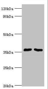
Figure 1. Western blot analysis of WDR77 using anti-WDR77 antibody (A04894-1). Electrophoresis was performed on a 5-20% SDS-PAGE gel at 70V (Stacking gel) / 90V (Resolving gel) for 2-3 hours. The sample well of each lane was loaded with 30 ug of sample under reducing conditions. Lane 1: human HEK293 whole cell lysates, Lane 2: human Hela whole cell lysates, Lane 3: human U20S whole cell lysates. After electrophoresis, proteins were transferred to a nitrocellulose membrane at 150 mA for 50-90 minutes. Blocked the membrane with 5% non-fat milk/TBS for 1.5 hour at RT. The membrane was incubated with rabbit anti-WDR77 antigen affinity purified polyclonal antibody (Catalog # A04894-1) at 0.25 microg/mL overnight at 4°C, then washed with TBS-0.1%Tween 3 times with 5 minutes each and probed with a goat anti-rabbit IgG-HRP secondary antibody at a dilution of 1:5000 for 1.5 hour at RT. The signal is developed using an Enhanced Chemiluminescent detection (ECL) kit (Catalog # EK1002) with Tanon 5200 system. A specific band was detected for WDR77 at approximately 42 kDa. The expected band size for WDR77 is at 42 kDa.
Figure 1. Western blot analysis of WDR77 using anti-WDR77 antibody (A04894-1). Electrophoresis was performed on a 5-20% SDS-PAGE gel at 70V (Stacking gel) / 90V (Resolving gel) for 2-3 hours. The sample well of each lane was loaded with 30 ug of sample under reducing conditions. Lane 1: human HEK293 whole cell lysates, Lane 2: human Hela whole cell lysates, Lane 3: human U20S whole cell lysates. After electrophoresis, proteins were transferred to a nitrocellulose membrane at 150 mA for 50-90 minutes. Blocked the membrane with 5% non-fat milk/TBS for 1.5 hour at RT. The membrane was incubated with rabbit anti-WDR77 antigen affinity purified polyclonal antibody (Catalog # A04894-1) at 0.25 microg/mL overnight at 4°C, then washed with TBS-0.1%Tween 3 times with 5 minutes each and probed with a goat anti-rabbit IgG-HRP secondary antibody at a dilution of 1:5000 for 1.5 hour at RT. The signal is developed using an Enhanced Chemiluminescent detection (ECL) kit (Catalog # EK1002) with Tanon 5200 system. A specific band was detected for WDR77 at approximately 42 kDa. The expected band size for WDR77 is at 42 kDa.
Anti-WDR77 Antibody Picoband(r)
A04894-1-CARRIER-FREE
ApplicationsWestern Blot, ELISA
Product group Antibodies
ReactivityHuman
TargetWDR77
Overview
- SupplierBoster Bio
- Product NameAnti-WDR77 Antibody Picoband(r)
- Delivery Days Customer9
- ApplicationsWestern Blot, ELISA
- CertificationResearch Use Only
- ClonalityPolyclonal
- Concentration500 ug/ml
- Gene ID79084
- Target nameWDR77
- Target descriptionWD repeat domain 77
- Target synonymsHKMT1069, MEP-50, MEP50, Nbla10071, p44, p44/Mep50, methylosome protein WDR77, WD repeat-containing protein 77, androgen receptor cofactor p44, methylosome protein 50, testis tissue sperm-binding protein Li 44a
- HostRabbit
- IsotypeIgG
- Protein IDQ9BQA1
- Protein NameMethylosome protein WDR77
- Scientific DescriptionBoster Bio Anti-WDR77 Antibody Picoband® catalog # A04894-1. Tested in ELISA, WB applications. This antibody reacts with Human. The brand Picoband indicates this is a premium antibody that guarantees superior quality, high affinity, and strong signals with minimal background in Western blot applications. Only our best-performing antibodies are designated as Picoband, ensuring unmatched performance.
- ReactivityHuman
- Storage Instruction-20°C,2°C to 8°C
- UNSPSC12352203
Related products
Product group Antibodies
Anti-WDR77 AntibodyA31898
ApplicationsImmunoFluorescence, Western Blot, ImmunoHistoChemistry
ReactivityHuman
- SizePrice
Product group Antibodies
Anti-WDR77 Antibody102-26561
ApplicationsWestern Blot, ImmunoCytoChemistry
TargetWDR77
- SizePrice
Product group Antibodies

WDR77 / MEP50 AntibodyLS-C831801
ApplicationsWestern Blot, ELISA
ReactivityHuman, Mouse
TargetWDR77
- SizePrice
Product group Antibodies
WDR77 AntibodyCSB-PA859030ESR2HU
ApplicationsWestern Blot, ELISA, ImmunoHistoChemistry
ReactivityHuman
TargetWDR77
- SizePrice

Product group Antibodies
Anti-WDR77 AntibodyHPA026437
ApplicationsImmunoHistoChemistry
ReactivityHuman
TargetWDR77
- SizePrice
Product group Antibodies
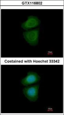
WDR77 antibody [N2C3]GTX116802
ApplicationsImmunoFluorescence, Western Blot, ImmunoCytoChemistry, ImmunoHistoChemistry, ImmunoHistoChemistry Paraffin
ReactivityHuman
TargetWDR77
- SizePrice
Product group Antibodies
Anti-WDR77 AntibodyCAB7134
ApplicationsImmunoFluorescence, Western Blot, ELISA, ImmunoCytoChemistry
ReactivityHuman
TargetWDR77
- SizePrice