


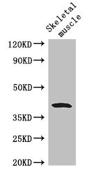
Figure 1. Western blot analysis of WISP1 using anti-WISP1 antibody (PA2089). Electrophoresis was performed on a 5-20% SDS-PAGE gel at 70V (Stacking gel) / 90V (Resolving gel) for 2-3 hours. The sample well of each lane was loaded with 30 ug of sample under reducing conditions. Lane 1: rat kidney tissue lysates, Lane 2: mouse kidney tissue lysates. After electrophoresis, proteins were transferred to a nitrocellulose membrane at 150 mA for 50-90 minutes. Blocked the membrane with 5% non-fat milk/TBS for 1.5 hour at RT. The membrane was incubated with rabbit anti-WISP1 antigen affinity purified polyclonal antibody (Catalog # PA2089) at 0.5 microg/mL overnight at 4°C, then washed with TBS-0.1%Tween 3 times with 5 minutes each and probed with a goat anti-rabbit IgG-HRP secondary antibody at a dilution of 1:5000 for 1.5 hour at RT. The signal is developed using an Enhanced Chemiluminescent detection (ECL) kit (Catalog # EK1002) with Tanon 5200 system. A specific band was detected for WISP1 at approximately 45 kDa. The expected band size for WISP1 is at 40 kDa.
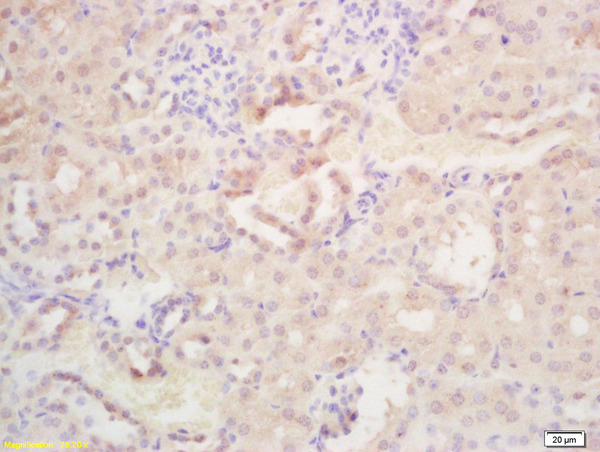
. WISP1 was detected in a paraffin-embedded section of human colorectal adenocarcinoma tissue. Heat mediated antigen retrieval was performed in EDTA buffer (pH 8.0, epitope retrieval solution). The tissue section was blocked with 10% goat serum. The tissue section was then incubated with 2 microg/ml rabbit anti-WISP1 Antibody (PA2089) overnight at 4°C. Peroxidase Conjugated Goat Anti-rabbit IgG was used as secondary antibody and incubated for 30 minutes at 37°C. The tissue section was developed using HRP Conjugated Rabbit IgG Super Vision Assay Kit (Catalog # SV0002) with DAB as the chromogen.")
. WISP1 was detected in a paraffin-embedded section of human testicular germ cell tumor tissue. Heat mediated antigen retrieval was performed in EDTA buffer (pH 8.0, epitope retrieval solution). The tissue section was blocked with 10% goat serum. The tissue section was then incubated with 2 microg/ml rabbit anti-WISP1 Antibody (PA2089) overnight at 4°C. Peroxidase Conjugated Goat Anti-rabbit IgG was used as secondary antibody and incubated for 30 minutes at 37°C. The tissue section was developed using HRP Conjugated Rabbit IgG Super Vision Assay Kit (Catalog # SV0002) with DAB as the chromogen.")
Figure 1. Western blot analysis of WISP1 using anti-WISP1 antibody (PA2089). Electrophoresis was performed on a 5-20% SDS-PAGE gel at 70V (Stacking gel) / 90V (Resolving gel) for 2-3 hours. The sample well of each lane was loaded with 30 ug of sample under reducing conditions. Lane 1: rat kidney tissue lysates, Lane 2: mouse kidney tissue lysates. After electrophoresis, proteins were transferred to a nitrocellulose membrane at 150 mA for 50-90 minutes. Blocked the membrane with 5% non-fat milk/TBS for 1.5 hour at RT. The membrane was incubated with rabbit anti-WISP1 antigen affinity purified polyclonal antibody (Catalog # PA2089) at 0.5 microg/mL overnight at 4°C, then washed with TBS-0.1%Tween 3 times with 5 minutes each and probed with a goat anti-rabbit IgG-HRP secondary antibody at a dilution of 1:5000 for 1.5 hour at RT. The signal is developed using an Enhanced Chemiluminescent detection (ECL) kit (Catalog # EK1002) with Tanon 5200 system. A specific band was detected for WISP1 at approximately 45 kDa. The expected band size for WISP1 is at 40 kDa.
Anti-WISP1 Antibody Picoband(r)
PA2089
ApplicationsWestern Blot, ImmunoHistoChemistry
Product group Antibodies
ReactivityBovine, Human, Mouse, Rat
TargetCCN4
Overview
- SupplierBoster Bio
- Product NameAnti-WISP1 Antibody Picoband(r)
- Delivery Days Customer9
- Application Supplier NoteTested Species: In-house tested species with positive results. Predicted Species: Species predicted to be fit for the product based on sequence similarities. By Heat: Boiling the paraffin sections in 10mM citrate buffer, pH6.0, for 20mins is required for the staining of formalin/paraffin sections. Other applications have not been tested. Optimal dilutions should be determined by end users.
- ApplicationsWestern Blot, ImmunoHistoChemistry
- Applications SupplierIHP, IHF, ICC, WB, IHC
- CertificationResearch Use Only
- ClonalityPolyclonal
- Concentration500 ug/ml
- Gene ID8840
- Target nameCCN4
- Target descriptioncellular communication network factor 4
- Target synonymsWISP1, WISP1-OT1, WISP1-UT1, WISP1c, WISP1i, WISP1tc, CCN family member 4, WISP1 3'UTR-associated RNA 1, WISP1 overlapping transcript 1 (non-protein coding), WNT1 induced secreted protein 1, WNT1 inducible signaling pathway protein 1
- HostRabbit
- IsotypeIgG
- Protein IDO95388
- Protein NameCCN family member 4
- Scientific DescriptionBoster Bio Anti-WISP1 Antibody catalog # PA2089. Tested in IHC, WB applications. This antibody reacts with Human, Mouse, Rat. The brand Picoband indicates this is a premium antibody that guarantees superior quality, high affinity, and strong signals with minimal background in Western blot applications. Only our best-performing antibodies are designated as Picoband, ensuring unmatched performance.
- ReactivityBovine, Human, Mouse, Rat
- Reactivity SupplierHuman, Mouse, Rat, Bovine
- Storage Instruction-20°C,2°C to 8°C
- UNSPSC12352203
References
- Liu L, Xu S, Li P, et al. A novel adipokine WISP1 attenuates lipopolysaccharide-induced cell injury in 3T3-L1 adipocytes by regulating the PI3K/Akt pathway. Obes Res Clin Pract. 2022,16(2):122-129. doi: 10.1016/j.orcp.2022.03.001Read this paper
- Yang R, Jia Q, Ma SF, et al. Exogenous H2S mitigates myocardial fibrosis in diabetic rats through suppression of the canonical Wnt pathway. Int J Mol Med. 2019,44(2):549-558. doi: 10.3892/ijmm.2019.4237Read this paper
Datasheet
MSDS
Related products
Product group Antibodies
WISP1 AntibodyCSB-PA026119LA01HU
ApplicationsImmunoFluorescence, Western Blot, ELISA, ImmunoHistoChemistry
ReactivityHuman, Mouse
TargetCCN4
- SizePrice
Product group Antibodies
Anti-WISP1 AntibodyA11637
ApplicationsWestern Blot
ReactivityHuman, Mouse
- SizePrice

Product group Antibodies
Anti-WISP-1/CCN4 Antibody Picoband(r)A03052-1-CARRIER-FREE
ApplicationsWestern Blot, ELISA
ReactivityHuman, Mouse, Rat
TargetCCN4
- SizePrice
Product group Antibodies

Goat anti-WISP1EB08178
ApplicationsFlow Cytometry, ImmunoFluorescence, Western Blot, ELISA
ReactivityHuman, Mouse, Rat
TargetCCN4
- SizePrice

Product group Antibodies
Anti-WISP1 AntibodyHPA007121
ApplicationsImmunoCytoChemistry
ReactivityHuman
TargetCCN4
- SizePrice
Product group Antibodies

CCN4 / WISP1 AntibodyLS-C332504
ApplicationsWestern Blot
ReactivityHuman, Mouse, Rat
TargetCCN4
- SizePrice
Product group Antibodies
WISP1 Polyclonal AntibodyBS-6321R
ApplicationsImmunoFluorescence, ELISA, ImmunoCytoChemistry, ImmunoHistoChemistry, ImmunoHistoChemistry Frozen, ImmunoHistoChemistry Paraffin
ReactivityHuman, Mouse, Rabbit, Rat
TargetCCN4
- SizePrice
Product group Antibodies
Wisp1 Polyclonal AntibodyCAC07598
ApplicationsImmunoFluorescence, Western Blot, ELISA, ImmunoHistoChemistry
ReactivityMouse
TargetCCN4
- SizePrice
Product group Antibodies
WISP1 antibody, C-termGTX88924
ApplicationsWestern Blot
ReactivityHuman
TargetCCN4
- SizePrice
Product group Antibodies
Anti-WISP1 (Center) Antibody102-26986
ApplicationsWestern Blot, ImmunoHistoChemistry, ImmunoHistoChemistry Paraffin
TargetCCN4
- SizePrice