


Figure 2. Flow Cytometry analysis of PC-3 cells using anti-WNT7A antibody (A01728). Overlay histogram showing PC-3 cells stained with A01728 (Blue line). The cells were fixed with 4% paraformaldehyde and blocked with 10% normal goat serum. And then incubated with rabbit anti-WNT7A Antibody (A01728, 1 microg/1x106 cells) for 30 min at 20°C. DyLight®488 conjugated goat anti-rabbit IgG (BA1127, 5-10 microg/1x106 cells) was used as secondary antibody for 30 minutes at 20°C. Isotype control antibody (Green line) was rabbit IgG (1 microg/1x106) used under the same conditions. Unlabelled sample without incubation with primary antibody and secondary antibody (Red line) was used as a blank control.
. Overlay histogram showing ANA-1 cells stained with A01728 (Blue line). The cells were fixed with 4% paraformaldehyde and blocked with 10% normal goat serum. And then incubated with rabbit anti-WNT7A Antibody (A01728, 1 microg/1x106 cells) for 30 min at 20°C. DyLight®488 conjugated goat anti-rabbit IgG (BA1127, 5-10 microg/1x106 cells) was used as secondary antibody for 30 minutes at 20°C. Isotype control antibody (Green line) was rabbit IgG (1 microg/1x106) used under the same conditions. Unlabelled sample without incubation with primary antibody and secondary antibody (Red line) was used as a blank control.")
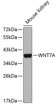
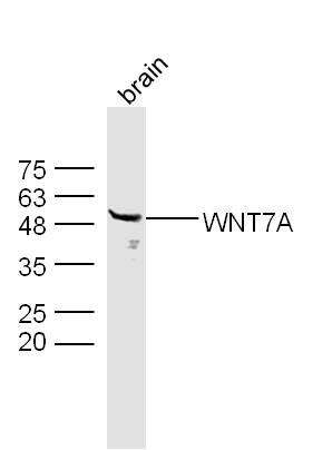
. Electrophoresis was performed on a 5-20% SDS-PAGE gel at 70V (Stacking gel) / 90V (Resolving gel) for 2-3 hours. The sample well of each lane was loaded with 30 ug of sample under reducing conditions. Lane 1: human hepatocellular carcinoma paracancerous tissue (HCCP) lysates, Lane 2: rat kidney tissue lysates, Lane 3: rat liver tissue lysates, Lane 4: mouse kidney tissue lysates, Lane 5: mouse liver tissue lysates. After electrophoresis, proteins were transferred to a nitrocellulose membrane at 150 mA for 50-90 minutes. Blocked the membrane with 5% non-fat milk/TBS for 1.5 hour at RT. The membrane was incubated with rabbit anti-WNT7A antigen affinity purified polyclonal antibody (Catalog # A01728) at 0.5 microg/mL overnight at 4°C, then washed with TBS-0.1%Tween 3 times with 5 minutes each and probed with a goat anti-rabbit IgG-DyLight 647 Conjugated secondary antibody at a dilution of 1:5000 for 1.5 hour at RT. A specific band was detected for WNT7A at approximately 40 kDa. The expected band size for WNT7A is at 40 kDa.")
Figure 2. Flow Cytometry analysis of PC-3 cells using anti-WNT7A antibody (A01728). Overlay histogram showing PC-3 cells stained with A01728 (Blue line). The cells were fixed with 4% paraformaldehyde and blocked with 10% normal goat serum. And then incubated with rabbit anti-WNT7A Antibody (A01728, 1 microg/1x106 cells) for 30 min at 20°C. DyLight®488 conjugated goat anti-rabbit IgG (BA1127, 5-10 microg/1x106 cells) was used as secondary antibody for 30 minutes at 20°C. Isotype control antibody (Green line) was rabbit IgG (1 microg/1x106) used under the same conditions. Unlabelled sample without incubation with primary antibody and secondary antibody (Red line) was used as a blank control.
Anti-WNT7A Antibody Picoband(r)
A01728-CARRIER-FREE
ApplicationsFlow Cytometry, Western Blot
Product group Antibodies
ReactivityHuman, Mouse, Rat
TargetWNT7A
Overview
- SupplierBoster Bio
- Product NameAnti-WNT7A Antibody Picoband(r)
- Delivery Days Customer9
- Application Supplier NoteTested Species: In-house tested species with positive results. Other applications have not been tested. Optimal dilutions should be determined by end users.
- ApplicationsFlow Cytometry, Western Blot
- CertificationResearch Use Only
- ClonalityPolyclonal
- Concentration500 ug/ml
- Gene ID7476
- Target nameWNT7A
- Target descriptionWnt family member 7A
- Target synonymsSANTOS, Wnt-7a, protein Wnt-7a, proto-oncogene Wnt7a protein, wingless-type MMTV integration site family, member 7A
- HostRabbit
- IsotypeIgG
- Protein IDO00755
- Protein NameProtein Wnt-7a
- Scientific DescriptionBoster Bio Anti-WNT7A Antibody Picoband® catalog # A01728. Tested in Flow Cytometry, WB applications. This antibody reacts with Human, Mouse, Rat. The brand Picoband indicates this is a premium antibody that guarantees superior quality, high affinity, and strong signals with minimal background in Western blot applications. Only our best-performing antibodies are designated as Picoband, ensuring unmatched performance.
- ReactivityHuman, Mouse, Rat
- Storage Instruction-20°C,2°C to 8°C
- UNSPSC12352203
Related products
Product group Antibodies
Anti-WNT7A Antibody144-65910
ApplicationsImmunoFluorescence, Western Blot
ReactivityHuman, Mouse
TargetWNT7A
- SizePrice
Product group Antibodies
Anti-Wnt7a AntibodyA14754
ApplicationsWestern Blot
ReactivityHuman, Mouse
- SizePrice
Product group Antibodies

WNT7A AntibodyLS-C749204
ApplicationsWestern Blot
ReactivityHuman, Mouse, Rat
TargetWNT7A
- SizePrice
Product group Antibodies
WNT7A Polyclonal AntibodyBS-6645R
ApplicationsWestern Blot, ELISA
ReactivityHuman, Mouse, Rat
TargetWNT7A
- SizePrice
Product group Antibodies
WNT7A AntibodyCSB-PA026141LA01HU
ApplicationsImmunoFluorescence, ELISA, ImmunoHistoChemistry
ReactivityHuman
TargetWNT7A
- SizePrice
Product group Antibodies
Wnt7a antibodyGTX105680
ApplicationsWestern Blot, ImmunoHistoChemistry, ImmunoHistoChemistry Paraffin
ReactivityHuman, Mouse
TargetWNT7A
- SizePrice

Product group Antibodies
Anti-WNT7A AntibodyHPA015719
ApplicationsImmunoHistoChemistry
ReactivityHuman
TargetWNT7A
- SizePrice
Product group Antibodies
Anti-WNT7A AntibodyCAB5425
ApplicationsWestern Blot, ELISA
ReactivityHuman
TargetWNT7A
- SizePrice