


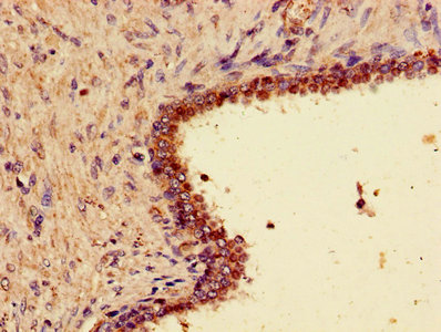
Immunohistochemical staining of human testis shows strong membrane and cytoplasmic positivity in cells in seminiferus ducts.
Immunohistochemical staining of human testis shows strong membrane and cytoplasmic positivity in cells in seminiferus ducts.
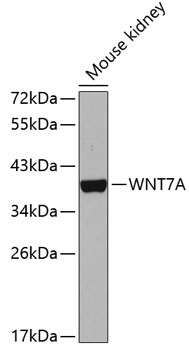
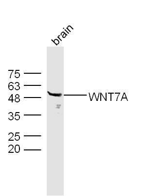
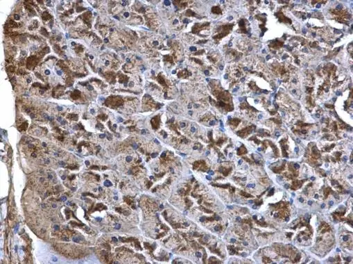
Anti-WNT7A Antibody
HPA015719
ApplicationsImmunoHistoChemistry
Product group Antibodies
ReactivityHuman
TargetWNT7A
Overview
- SupplierAtlas Antibodies
- Product NameAnti-WNT7A Antibody
- Delivery Days Customer4
- ApplicationsImmunoHistoChemistry
- CertificationResearch Use Only
- ClonalityPolyclonal
- ConjugateUnconjugated
- Gene ID7476
- Target nameWNT7A
- Target descriptionWnt family member 7A
- Target synonymsSANTOS, Wnt-7a, protein Wnt-7a, proto-oncogene Wnt7a protein, wingless-type MMTV integration site family, member 7A
- HostRabbit
- IsotypeIgG
- Protein IDO00755
- Protein NameProtein Wnt-7a
- Scientific DescriptionRecombinant Protein Epitope Signature Tag (PrEST) antigen sequence
- ReactivityHuman
- Storage Instruction-20°C,2°C to 8°C
- UNSPSC41116161
Datasheet
MSDS
Related products
Product group Antibodies
Anti-WNT7A Antibody144-65910
ApplicationsImmunoFluorescence, Western Blot
ReactivityHuman, Mouse
TargetWNT7A
- SizePrice
Product group Antibodies
Anti-Wnt7a AntibodyA14754
ApplicationsWestern Blot
ReactivityHuman, Mouse
- SizePrice
Product group Antibodies

WNT7A AntibodyLS-C749204
ApplicationsWestern Blot
ReactivityHuman, Mouse, Rat
TargetWNT7A
- SizePrice

Product group Antibodies
Anti-WNT7A Antibody Picoband(r)A01728-CARRIER-FREE
ApplicationsFlow Cytometry, Western Blot
ReactivityHuman, Mouse, Rat
TargetWNT7A
- SizePrice
Product group Antibodies
WNT7A Polyclonal AntibodyBS-6645R
ApplicationsWestern Blot, ELISA
ReactivityHuman, Mouse, Rat
TargetWNT7A
- SizePrice
Product group Antibodies
WNT7A AntibodyCSB-PA026141LA01HU
ApplicationsImmunoFluorescence, ELISA, ImmunoHistoChemistry
ReactivityHuman
TargetWNT7A
- SizePrice
Product group Antibodies
Wnt7a antibodyGTX105680
ApplicationsWestern Blot, ImmunoHistoChemistry, ImmunoHistoChemistry Paraffin
ReactivityHuman, Mouse
TargetWNT7A
- SizePrice
Product group Antibodies
Anti-WNT7A AntibodyCAB5425
ApplicationsWestern Blot, ELISA
ReactivityHuman
TargetWNT7A
- SizePrice