


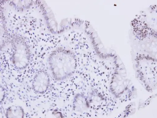
Immunohistochemical staining of human testis shows moderate nuclear positivity in Leydig cells.

Immunohistochemical staining of human testis shows moderate nuclear positivity in Leydig cells.
Anti-WRN Antibody
HPA028661
ApplicationsImmunoHistoChemistry
Product group Antibodies
ReactivityHuman
TargetWRN
Overview
- SupplierAtlas Antibodies
- Product NameAnti-WRN Antibody
- Delivery Days Customer4
- ApplicationsImmunoHistoChemistry
- CertificationResearch Use Only
- ClonalityPolyclonal
- ConjugateUnconjugated
- Gene ID7486
- Target nameWRN
- Target descriptionWRN RecQ like helicase
- Target synonymsRECQ3, RECQL2, RECQL3, bifunctional 3'-5' exonuclease/ATP-dependent helicase WRN, DNA helicase, RecQ-like type 3, Werner syndrome ATP-dependent helicase, Werner syndrome RecQ like helicase, Werner syndrome protein, Werner syndrome, RecQ helicase-like, exonuclease WRN, recQ protein-like 2
- HostRabbit
- IsotypeIgG
- Protein IDQ14191
- Protein NameBifunctional 3'-5' exonuclease/ATP-dependent helicase WRN
- Scientific DescriptionRecombinant Protein Epitope Signature Tag (PrEST) antigen sequence
- ReactivityHuman
- Storage Instruction-20°C,2°C to 8°C
- UNSPSC41116161
Datasheet
MSDS
Related products
Product group Antibodies
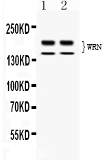
Anti-WRN Antibody144-06855
ApplicationsWestern Blot
ReactivityHuman
TargetWRN
- SizePrice
Product group Antibodies
WRN Recombinant AntibodyBSM-54700R
ApplicationsWestern Blot
ReactivityHuman
TargetWRN
- SizePrice
Product group Antibodies
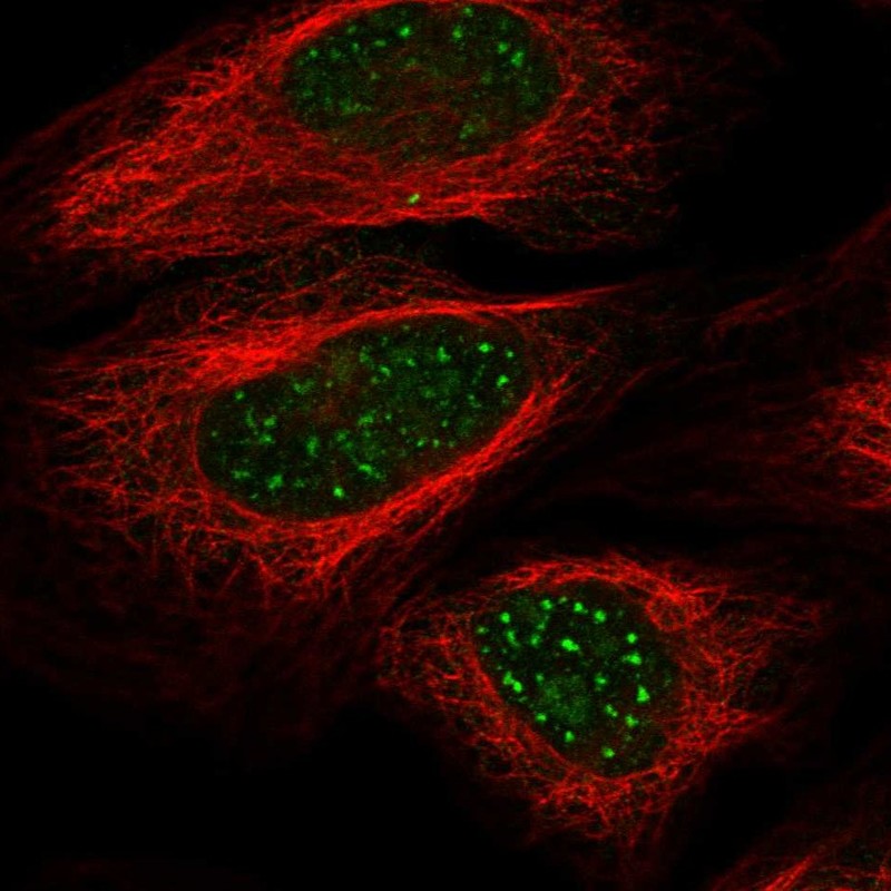
Anti-WRN AntibodyHPA009423
ApplicationsImmunoCytoChemistry
ReactivityHuman
TargetWRN
- SizePrice
Product group Antibodies
Phospho-WRN (S1141) AntibodyCSB-PA060086
ApplicationsWestern Blot, ELISA
ReactivityHuman
TargetWRN
- SizePrice
Product group Antibodies
References
WRN antibodyGTX101081
ApplicationsImmunoFluorescence, ImmunoPrecipitation, Western Blot, ImmunoCytoChemistry, ImmunoHistoChemistry, ImmunoHistoChemistry Paraffin
ReactivityHuman
TargetWRN
- SizePrice
Product group Antibodies
Anti-Werners syndrome helicase WRN/WRN Antibody Picoband(r)PB10107-CARRIER-FREE
ApplicationsWestern Blot
ReactivityHuman, Rat
TargetWRN
- SizePrice
Product group Antibodies
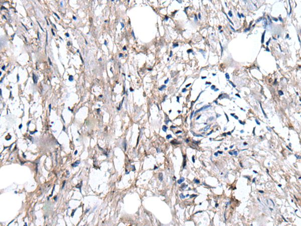
Anti-WRN AntibodyA47278
ApplicationsImmunoHistoChemistry
ReactivityHuman
- SizePrice