


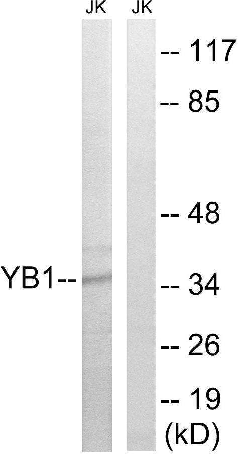
Figure 1. Western blot analysis of YBX1 using anti-YBX1 antibody (PA1758). Electrophoresis was performed on a 5-20% SDS-PAGE gel at 70V (Stacking gel) / 90V (Resolving gel) for 2-3 hours. The sample well of each lane was loaded with 30 ug of sample under reducing conditions. Lane 1: human MCF-7 whole cell lysates, Lane 2: human 293T whole cell lysates, Lane 3: human Hela whole cell lysates, Lane 4: human Jurkat whole cell lysates, Lane 5: human T47D whole cell lysates, Lane 6: human THP-1 whole cell lysates, Lane 7: human MOLT4 whole cell lysates, Lane 8: human HL-60 whole cell lysates, Lane 9: rat testis tissue lysates, Lane 10: mouse stomach tissue lysates, Lane 11: mouse testis tissue lysates. After electrophoresis, proteins were transferred to a nitrocellulose membrane at 150 mA for 50-90 minutes. Blocked the membrane with 5% non-fat milk/TBS for 1.5 hour at RT. The membrane was incubated with rabbit anti-YBX1 antigen affinity purified polyclonal antibody (Catalog # PA1758) at 0.5 microg/mL overnight at 4°C, then washed with TBS-0.1%Tween 3 times with 5 minutes each and probed with a goat anti-rabbit IgG-HRP secondary antibody at a dilution of 1:5000 for 1.5 hour at RT. The signal is developed using an Enhanced Chemiluminescent detection (ECL) kit (Catalog # EK1002) with Tanon 5200 system. A specific band was detected for YBX1 at approximately 50 kDa. The expected band size for YBX1 is at 36 kDa.
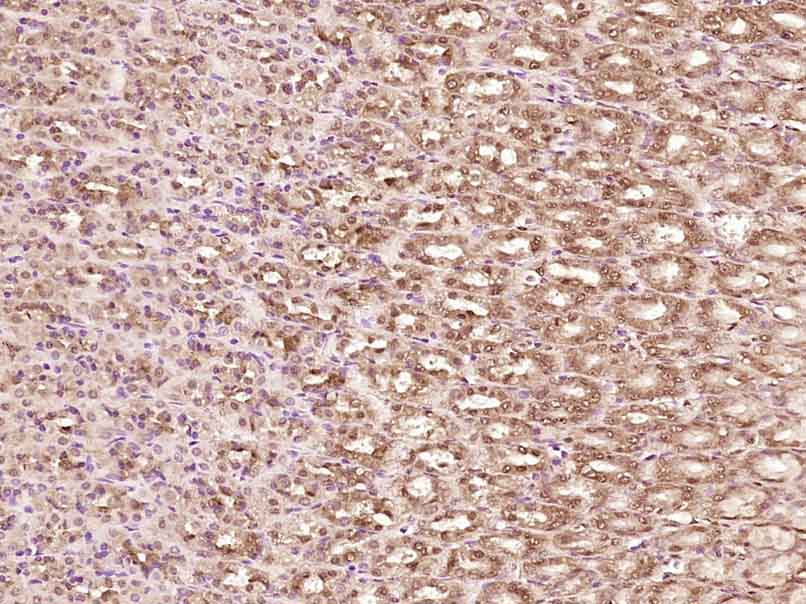
. YBX1 was detected in a paraffin-embedded section of human rectal cancer tissue. Heat mediated antigen retrieval was performed in EDTA buffer (pH 8.0, epitope retrieval solution). The tissue section was blocked with 10% goat serum. The tissue section was then incubated with 2 microg/ml rabbit anti-YBX1 Antibody (PA1758) overnight at 4°C. Peroxidase Conjugated Goat Anti-rabbit IgG was used as secondary antibody and incubated for 30 minutes at 37°C. The tissue section was developed using HRP Conjugated Rabbit IgG Super Vision Assay Kit (Catalog # SV0002) with DAB as the chromogen.")
. YBX1 was detected in a paraffin-embedded section of human ovarian cancer tissue. Heat mediated antigen retrieval was performed in EDTA buffer (pH 8.0, epitope retrieval solution). The tissue section was blocked with 10% goat serum. The tissue section was then incubated with 2 microg/ml rabbit anti-YBX1 Antibody (PA1758) overnight at 4°C. Peroxidase Conjugated Goat Anti-rabbit IgG was used as secondary antibody and incubated for 30 minutes at 37°C. The tissue section was developed using HRP Conjugated Rabbit IgG Super Vision Assay Kit (Catalog # SV0002) with DAB as the chromogen.")
. YBX1 was detected in a paraffin-embedded section of mouse testis tissue. Heat mediated antigen retrieval was performed in EDTA buffer (pH 8.0, epitope retrieval solution). The tissue section was blocked with 10% goat serum. The tissue section was then incubated with 2 microg/ml rabbit anti-YBX1 Antibody (PA1758) overnight at 4°C. Peroxidase Conjugated Goat Anti-rabbit IgG was used as secondary antibody and incubated for 30 minutes at 37°C. The tissue section was developed using HRP Conjugated Rabbit IgG Super Vision Assay Kit (Catalog # SV0002) with DAB as the chromogen.")
. YBX1 was detected in a paraffin-embedded section of rat testis tissue. Heat mediated antigen retrieval was performed in EDTA buffer (pH 8.0, epitope retrieval solution). The tissue section was blocked with 10% goat serum. The tissue section was then incubated with 2 microg/ml rabbit anti-YBX1 Antibody (PA1758) overnight at 4°C. Peroxidase Conjugated Goat Anti-rabbit IgG was used as secondary antibody and incubated for 30 minutes at 37°C. The tissue section was developed using HRP Conjugated Rabbit IgG Super Vision Assay Kit (Catalog # SV0002) with DAB as the chromogen.")
. Overlay histogram showing HEL cells stained with PA1758 (Blue line). To facilitate intracellular staining, cells were fixed with 4% paraformaldehyde and permeabilized with permeabilization buffer. The cells were blocked with 10% normal goat serum. And then incubated with rabbit anti-YBX1 Antibody (PA1758, 1 microg/1x106 cells) for 30 min at 20°C. DyLight®488 conjugated goat anti-rabbit IgG (BA1127, 5-10 microg/1x106 cells) was used as secondary antibody for 30 minutes at 20°C. Isotype control antibody (Green line) was rabbit IgG (1 microg/1x106) used under the same conditions. Unlabelled sample without incubation with primary antibody and secondary antibody (Red line) was used as a blank control.")
Figure 1. Western blot analysis of YBX1 using anti-YBX1 antibody (PA1758). Electrophoresis was performed on a 5-20% SDS-PAGE gel at 70V (Stacking gel) / 90V (Resolving gel) for 2-3 hours. The sample well of each lane was loaded with 30 ug of sample under reducing conditions. Lane 1: human MCF-7 whole cell lysates, Lane 2: human 293T whole cell lysates, Lane 3: human Hela whole cell lysates, Lane 4: human Jurkat whole cell lysates, Lane 5: human T47D whole cell lysates, Lane 6: human THP-1 whole cell lysates, Lane 7: human MOLT4 whole cell lysates, Lane 8: human HL-60 whole cell lysates, Lane 9: rat testis tissue lysates, Lane 10: mouse stomach tissue lysates, Lane 11: mouse testis tissue lysates. After electrophoresis, proteins were transferred to a nitrocellulose membrane at 150 mA for 50-90 minutes. Blocked the membrane with 5% non-fat milk/TBS for 1.5 hour at RT. The membrane was incubated with rabbit anti-YBX1 antigen affinity purified polyclonal antibody (Catalog # PA1758) at 0.5 microg/mL overnight at 4°C, then washed with TBS-0.1%Tween 3 times with 5 minutes each and probed with a goat anti-rabbit IgG-HRP secondary antibody at a dilution of 1:5000 for 1.5 hour at RT. The signal is developed using an Enhanced Chemiluminescent detection (ECL) kit (Catalog # EK1002) with Tanon 5200 system. A specific band was detected for YBX1 at approximately 50 kDa. The expected band size for YBX1 is at 36 kDa.
Anti-YB1/YBX1 Antibody Picoband(r)
PA1758
ApplicationsFlow Cytometry, Western Blot, ImmunoHistoChemistry
Product group Antibodies
ReactivityHamster, Human, Mouse, Rat
TargetYBX1
Overview
- SupplierBoster Bio
- Product NameAnti-YB1/YBX1 Antibody Picoband(r)
- Delivery Days Customer9
- Application Supplier NoteTested Species: In-house tested species with positive results. Predicted Species: Species predicted to be fit for the product based on sequence similarities. Other applications have not been tested. Optimal dilutions should be determined by end users.
- ApplicationsFlow Cytometry, Western Blot, ImmunoHistoChemistry
- Applications SupplierWB
- CertificationResearch Use Only
- ClonalityPolyclonal
- Concentration500 ug/ml
- Gene ID4904
- Target nameYBX1
- Target descriptionY-box binding protein 1
- Target synonymsBP-8, CBF-A, CSDA2, CSDB, DBPB, EFI-A, MDR-NF1, NSEP-1, NSEP1, YB-1, YB1, Y-box-binding protein 1, CCAAT-binding transcription factor I subunit A, DNA-binding protein B, Y-box transcription factor, enhancer factor I subunit A, major histocompatibility complex, class II, Y box-binding protein I, nuclease-sensitive element-binding protein 1
- HostRabbit
- IsotypeIgG
- Protein IDP67809
- Protein NameY-box-binding protein 1
- Scientific DescriptionBoster Bio Anti-YB1/YBX1 Antibody catalog # PA1758. Tested in Flow Cytometry, IHC, WB applications. This antibody reacts with Human, Mouse, Rat. The brand Picoband indicates this is a premium antibody that guarantees superior quality, high affinity, and strong signals with minimal background in Western blot applications. Only our best-performing antibodies are designated as Picoband, ensuring unmatched performance.
- ReactivityHamster, Human, Mouse, Rat
- Reactivity SupplierHuman, Mouse, Rat, Hamster
- Storage Instruction-20°C,2°C to 8°C
- UNSPSC12352203
Datasheet
MSDS
Related products
Product group Antibodies
Phospho-YBX1 (S102) AntibodyCSB-PA020157
ApplicationsWestern Blot, ELISA
ReactivityHuman, Mouse, Rat
TargetYBX1
- SizePrice
Product group Antibodies
Anti-YB1 AntibodyA98415
ApplicationsWestern Blot, ELISA
ReactivityHuman, Mouse, Rat
- SizePrice
Product group Antibodies

YBX1 / YB1 AntibodyLS-C832725
ApplicationsELISA, ImmunoHistoChemistry
ReactivityHuman, Mouse, Rat
TargetYBX1
- SizePrice

Product group Antibodies
Anti-YBX1 AntibodyHPA040304
ApplicationsWestern Blot, ImmunoCytoChemistry, ImmunoHistoChemistry
ReactivityHuman, Mouse, Rat
TargetYBX1
- SizePrice
Product group Antibodies
ApplicationsImmunoHistoChemistry
ReactivityPorcine
TargetYBX1
- SizePrice

Product group Antibodies
Anti-YB1/YBX1 Antibody Picoband(r)PB9465-CARRIER-FREE
ApplicationsFlow Cytometry, ImmunoFluorescence, Western Blot, ImmunoCytoChemistry, ImmunoHistoChemistry, ImmunoHistoChemistry Frozen
ReactivityHamster, Human, Mouse, Rat
TargetYBX1
- SizePrice
Product group Antibodies
ApplicationsImmunoFluorescence, Western Blot, ELISA, ImmunoCytoChemistry, ImmunoHistoChemistry, ImmunoHistoChemistry Frozen, ImmunoHistoChemistry Paraffin
ReactivityBovine, Canine, Chicken, Equine, Guinea Pig, Human, Mouse, Porcine, Rabbit, Rat
TargetYBX1
- SizePrice
Product group Antibodies
YB1 antibodyGTX131630
ApplicationsWestern Blot
ReactivityHuman
TargetYBX1
- SizePrice