


Figure 1. Western blot analysis of ZAP70 using anti-ZAP70 antibody (PB9968). Electrophoresis was performed on a 5-20% SDS-PAGE gel at 70V (Stacking gel) / 90V (Resolving gel) for 2-3 hours. The sample well of each lane was loaded with 30 ug of sample under reducing conditions. Lane 1: human Jurkat whole cell lysates, Lane 2: human CCRF-CEM whole cell lysates, Lane 3: human MOLT-4 whole cell lysates, Lane 4: human Raji whole cell lysates, Lane 5: rat spleen tissue lysates, Lane 6: rat thymus tissue lysates, Lane 7: mouse spleen tissue lysates, Lane 8: mouse thymus tissue lysates. After electrophoresis, proteins were transferred to a nitrocellulose membrane at 150 mA for 50-90 minutes. Blocked the membrane with 5% non-fat milk/TBS for 1.5 hour at RT. The membrane was incubated with rabbit anti-ZAP70 antigen affinity purified polyclonal antibody (Catalog # PB9968) at 0.5 microg/mL overnight at 4°C, then washed with TBS-0.1%Tween 3 times with 5 minutes each and probed with a goat anti-rabbit IgG-HRP secondary antibody at a dilution of 1:5000 for 1.5 hour at RT. The signal is developed using an Enhanced Chemiluminescent detection (ECL) kit (Catalog # EK1002) with Tanon 5200 system. A specific band was detected for ZAP70 at approximately 70 kDa. The expected band size for ZAP70 is at 70 kDa.
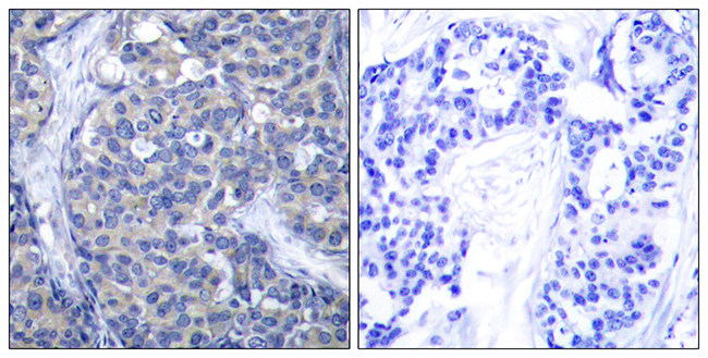
. ZAP70 was detected in a paraffin-embedded section of human prostate cancer tissue. Heat mediated antigen retrieval was performed in EDTA buffer (pH 8.0, epitope retrieval solution). The tissue section was blocked with 10% goat serum. The tissue section was then incubated with 2 microg/ml rabbit anti-ZAP70 Antibody (PB9968) overnight at 4°C. Peroxidase Conjugated Goat Anti-rabbit IgG was used as secondary antibody and incubated for 30 minutes at 37°C. The tissue section was developed using HRP Conjugated Rabbit IgG Super Vision Assay Kit (Catalog # SV0002) with DAB as the chromogen.")
Figure 1. Western blot analysis of ZAP70 using anti-ZAP70 antibody (PB9968). Electrophoresis was performed on a 5-20% SDS-PAGE gel at 70V (Stacking gel) / 90V (Resolving gel) for 2-3 hours. The sample well of each lane was loaded with 30 ug of sample under reducing conditions. Lane 1: human Jurkat whole cell lysates, Lane 2: human CCRF-CEM whole cell lysates, Lane 3: human MOLT-4 whole cell lysates, Lane 4: human Raji whole cell lysates, Lane 5: rat spleen tissue lysates, Lane 6: rat thymus tissue lysates, Lane 7: mouse spleen tissue lysates, Lane 8: mouse thymus tissue lysates. After electrophoresis, proteins were transferred to a nitrocellulose membrane at 150 mA for 50-90 minutes. Blocked the membrane with 5% non-fat milk/TBS for 1.5 hour at RT. The membrane was incubated with rabbit anti-ZAP70 antigen affinity purified polyclonal antibody (Catalog # PB9968) at 0.5 microg/mL overnight at 4°C, then washed with TBS-0.1%Tween 3 times with 5 minutes each and probed with a goat anti-rabbit IgG-HRP secondary antibody at a dilution of 1:5000 for 1.5 hour at RT. The signal is developed using an Enhanced Chemiluminescent detection (ECL) kit (Catalog # EK1002) with Tanon 5200 system. A specific band was detected for ZAP70 at approximately 70 kDa. The expected band size for ZAP70 is at 70 kDa.
Anti-ZAP70 Antibody Picoband(r)
PB9968-CARRIER-FREE
ApplicationsWestern Blot, ImmunoHistoChemistry
Product group Antibodies
ReactivityHuman, Mouse, Rat
TargetZAP70
Overview
- SupplierBoster Bio
- Product NameAnti-ZAP70 Antibody Picoband(r)
- Delivery Days Customer9
- Application Supplier NoteTested Species: In-house tested species with positive results. By Heat: Boiling the paraffin sections in 10mM citrate buffer, pH6.0, for 20mins is required for the staining of formalin/paraffin sections. Other applications have not been tested. Optimal dilutions should be determined by end users.
- ApplicationsWestern Blot, ImmunoHistoChemistry
- CertificationResearch Use Only
- ClonalityPolyclonal
- Concentration500 ug/ml
- Gene ID7535
- Target nameZAP70
- Target descriptionzeta chain of T cell receptor associated protein kinase 70
- Target synonymsADMIO2, IMD48, SRK, STCD, STD, TZK, ZAP-70, tyrosine-protein kinase ZAP-70, 70 kDa zeta-associated protein, 70 kDa zeta-chain associated protein, syk-related tyrosine kinase, zeta chain of T cell receptor associated protein kinase 70kDa, zeta-chain (TCR) associated protein kinase 70kDa, zeta-chain associated protein kinase, 70kD
- HostRabbit
- IsotypeIgG
- Protein IDP43403
- Protein NameTyrosine-protein kinase ZAP-70
- Scientific DescriptionBoster Bio Anti-ZAP70 Antibody Picoband® catalog # PB9968. Tested in IHC, WB applications. This antibody reacts with Human, Mouse, Rat. The brand Picoband indicates this is a premium antibody that guarantees superior quality, high affinity, and strong signals with minimal background in Western blot applications. Only our best-performing antibodies are designated as Picoband, ensuring unmatched performance.
- ReactivityHuman, Mouse, Rat
- Storage Instruction-20°C,2°C to 8°C
- UNSPSC12352203
Related products
Product group Antibodies
Phospho-ZAP70 (Y319) AntibodyCSB-PA060096
ApplicationsImmunoFluorescence, Western Blot, ELISA, ImmunoHistoChemistry
ReactivityHuman, Mouse, Rat
TargetZAP70
- SizePrice
Product group Antibodies
Anti-ZAP-70 AntibodyA95578
ApplicationsWestern Blot, ELISA, ImmunoHistoChemistry
ReactivityHuman, Mouse, Rat
- SizePrice
Product group Antibodies

ZAP70 AntibodyLS-C830821
ApplicationsWestern Blot, ELISA, ImmunoHistoChemistry
ReactivityHuman, Mouse
TargetZAP70
- SizePrice
Product group Antibodies
References

Goat anti-ZAP70EB11394
ApplicationsImmunoPrecipitation, Western Blot, ELISA
ReactivityBovine, Canine, Human, Mouse, Rat
TargetZAP70
- SizePrice

Product group Antibodies
Anti-ZAP70 AntibodyHPA003134
ApplicationsWestern Blot, ImmunoHistoChemistry
ReactivityHuman
TargetZAP70
- SizePrice
Product group Antibodies
ApplicationsImmunoPrecipitation, Western Blot, ImmunoCytoChemistry, ImmunoHistoChemistry
ReactivityPorcine
TargetZAP70
- SizePrice
Product group Antibodies
ZAP70 antibodyGTX101076
ApplicationsImmunoPrecipitation, Western Blot, ImmunoHistoChemistry, ImmunoHistoChemistry Paraffin
ReactivityHuman, Mouse
TargetZAP70
- SizePrice