


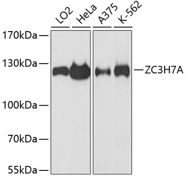
Figure 1. Western blot analysis of ZC3H7A using anti-ZC3H7A antibody (A15075-1). Electrophoresis was performed on a 5-20% SDS-PAGE gel at 70V (Stacking gel) / 90V (Resolving gel) for 2-3 hours. The sample well of each lane was loaded with 30 ug of sample under reducing conditions. Lane 1: human K562 whole cell lysates. After electrophoresis, proteins were transferred to a nitrocellulose membrane at 150 mA for 50-90 minutes. Blocked the membrane with 5% non-fat milk/TBS for 1.5 hour at RT. The membrane was incubated with rabbit anti-ZC3H7A antigen affinity purified polyclonal antibody (Catalog # A15075-1) at 0.5 microg/mL overnight at 4°C, then washed with TBS-0.1%Tween 3 times with 5 minutes each and probed with a goat anti-rabbit IgG-HRP secondary antibody at a dilution of 1:5000 for 1.5 hour at RT. The signal is developed using an Enhanced Chemiluminescent detection (ECL) kit (Catalog # EK1002) with Tanon 5200 system. A specific band was detected for ZC3H7A at approximately 111 kDa. The expected band size for ZC3H7A is at 111 kDa.
. ZC3H7A was detected in an immunocytochemical section of SiHa cells. Enzyme antigen retrieval was performed using IHC enzyme antigen retrieval reagent (AR0022) for 15 mins. The cells were blocked with 10% goat serum. And then incubated with 5 microg/mL rabbit anti-ZC3H7A Antibody (A15075-1) overnight at 4°C. DyLight®488 Conjugated Goat Anti-Rabbit IgG (BA1127) was used as secondary antibody at 1:100 dilution and incubated for 30 minutes at 37°C. The section was counterstained with DAPI. Visualize using a fluorescence microscope and filter sets appropriate for the label used.")
. Overlay histogram showing HL-60 cells stained with A15075-1 (Blue line). To facilitate intracellular staining, cells were fixed with 4% paraformaldehyde and permeabilized with permeabilization buffer. The cells were blocked with 10% normal goat serum. And then incubated with rabbit anti-ZC3H7A Antibody (A15075-1, 1 microg/1x106 cells) for 30 min at 20°C. DyLight®488 conjugated goat anti-rabbit IgG (BA1127, 5-10 microg/1x106 cells) was used as secondary antibody for 30 minutes at 20°C. Isotype control antibody (Green line) was rabbit IgG (1 microg/1x106) used under the same conditions. Unlabelled sample without incubation with primary antibody and secondary antibody (Red line) was used as a blank control.")
Figure 1. Western blot analysis of ZC3H7A using anti-ZC3H7A antibody (A15075-1). Electrophoresis was performed on a 5-20% SDS-PAGE gel at 70V (Stacking gel) / 90V (Resolving gel) for 2-3 hours. The sample well of each lane was loaded with 30 ug of sample under reducing conditions. Lane 1: human K562 whole cell lysates. After electrophoresis, proteins were transferred to a nitrocellulose membrane at 150 mA for 50-90 minutes. Blocked the membrane with 5% non-fat milk/TBS for 1.5 hour at RT. The membrane was incubated with rabbit anti-ZC3H7A antigen affinity purified polyclonal antibody (Catalog # A15075-1) at 0.5 microg/mL overnight at 4°C, then washed with TBS-0.1%Tween 3 times with 5 minutes each and probed with a goat anti-rabbit IgG-HRP secondary antibody at a dilution of 1:5000 for 1.5 hour at RT. The signal is developed using an Enhanced Chemiluminescent detection (ECL) kit (Catalog # EK1002) with Tanon 5200 system. A specific band was detected for ZC3H7A at approximately 111 kDa. The expected band size for ZC3H7A is at 111 kDa.
Anti-ZC3H7A Antibody Picoband(r)
A15075-1-CARRIER-FREE
ApplicationsFlow Cytometry, ImmunoFluorescence, Western Blot, ELISA, ImmunoCytoChemistry
Product group Antibodies
ReactivityHuman
TargetZC3H7A
Overview
- SupplierBoster Bio
- Product NameAnti-ZC3H7A Antibody Picoband(r)
- Delivery Days Customer9
- ApplicationsFlow Cytometry, ImmunoFluorescence, Western Blot, ELISA, ImmunoCytoChemistry
- CertificationResearch Use Only
- ClonalityPolyclonal
- Concentration500 ug/ml
- Gene ID29066
- Target nameZC3H7A
- Target descriptionzinc finger CCCH-type containing 7A
- Target synonymsHSPC055, ZC3H7, ZC3HDC7, zinc finger CCCH domain-containing protein 7A, zinc finger CCCH-type domain containing 7, zinc-finger protein AY163807
- HostRabbit
- IsotypeIgG
- Protein IDQ8IWR0
- Protein NameZinc finger CCCH domain-containing protein 7A
- Scientific DescriptionBoster Bio Anti-ZC3H7A Antibody Picoband® catalog # A15075-1. Tested in ELISA, Flow Cytometry, IF, ICC, WB applications. This antibody reacts with Human. The brand Picoband indicates this is a premium antibody that guarantees superior quality, high affinity, and strong signals with minimal background in Western blot applications. Only our best-performing antibodies are designated as Picoband, ensuring unmatched performance.
- ReactivityHuman
- Storage Instruction-20°C,2°C to 8°C
- UNSPSC12352203
Related products
Product group Antibodies
Anti-ZC3H7A AntibodyA88034
ApplicationsImmunoFluorescence, Western Blot, ImmunoCytoChemistry
ReactivityHuman, Mouse, Rat
- SizePrice
Product group Antibodies
Anti-ZC3H7A Antibody144-60230
ApplicationsWestern Blot
ReactivityHuman
TargetZC3H7A
- SizePrice

Product group Antibodies

ZC3H7A AntibodyLS-C832073
ApplicationsELISA, ImmunoHistoChemistry
ReactivityHuman
TargetZC3H7A
- SizePrice
Product group Antibodies
ZC3H7A AntibodyCSB-PA818245LA01HU
ApplicationsELISA, ImmunoHistoChemistry
ReactivityHuman
TargetZC3H7A
- SizePrice

Product group Antibodies
Anti-ZC3H7A AntibodyHPA040808
ApplicationsWestern Blot, ImmunoCytoChemistry, ImmunoHistoChemistry
ReactivityHuman
TargetZC3H7A
- SizePrice
Product group Antibodies
Anti-ZC3H7A AntibodyCAB13190
ApplicationsImmunoFluorescence, Western Blot, ELISA, ImmunoCytoChemistry
ReactivityHuman
TargetZC3H7A
- SizePrice