


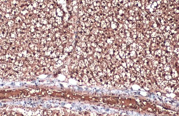
Immunohistochemical staining of human cerebral cortex shows strong nuclear positivity in neurons.

Immunohistochemical staining of human cerebral cortex shows strong nuclear positivity in neurons.
Anti-ZIC1 Antibody
HPA004098
ApplicationsImmunoCytoChemistry, ImmunoHistoChemistry
Product group Antibodies
ReactivityHuman
TargetZIC1
Overview
- SupplierAtlas Antibodies
- Product NameAnti-ZIC1 Antibody
- Delivery Days Customer4
- ApplicationsImmunoCytoChemistry, ImmunoHistoChemistry
- CertificationResearch Use Only
- ClonalityPolyclonal
- ConjugateUnconjugated
- Gene ID7545
- Target nameZIC1
- Target descriptionZic family zinc finger 1
- Target synonymsBAIDCS, CRS6, ZIC, ZNF201, zinc finger protein ZIC 1, Zic family member 1 (odd-paired homolog, Drosophila), Zinc finger protein of the cerebellum 1, zinc finger protein 201
- HostRabbit
- IsotypeIgG
- Protein IDQ15915
- Protein NameZinc finger protein ZIC 1
- Scientific DescriptionRecombinant Protein Epitope Signature Tag (PrEST) antigen sequence
- ReactivityHuman
- Storage Instruction-20°C,2°C to 8°C
- UNSPSC41116161
Datasheet
MSDS
Related products

Product group Antibodies
Anti-ZIC1 Antibody Picoband(r)A05537-4-CARRIER-FREE
ApplicationsWestern Blot, ELISA
ReactivityHuman, Mouse, Rat
TargetZIC1
- SizePrice
Product group Antibodies
ApplicationsImmunoFluorescence, Western Blot, ImmunoHistoChemistry
ReactivityHuman, Mouse, Rat
- SizePrice
Product group Antibodies
Anti-ZIC1 Antibody144-64995
ApplicationsWestern Blot
ReactivityHuman, Mouse, Rat
TargetZIC1
- SizePrice
Product group Antibodies

ZIC / ZIC1 AntibodyLS-C831137
ApplicationsELISA, ImmunoHistoChemistry
ReactivityHuman, Mouse
TargetZIC1
- SizePrice
Product group Antibodies
Zic1 Recombinant Antibody, AbBy Fluor-350 ConjugatedBSM-61588R-BF350
ApplicationsFlow Cytometry, ImmunoFluorescence, Western Blot, ImmunoCytoChemistry
ReactivityHuman, Mouse, Rat
TargetZIC1
- SizePrice
Product group Antibodies
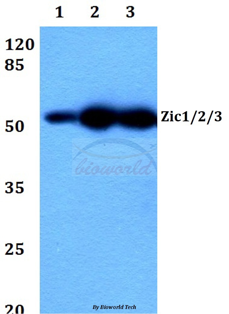
ZIC1/ZIC2/ZIC3 AntibodyCSB-PA060106
ApplicationsImmunoFluorescence, Western Blot, ELISA, ImmunoHistoChemistry
ReactivityHuman, Mouse
TargetZIC1
- SizePrice
Product group Antibodies
ZIC1 antibodyGTX129468
ApplicationsWestern Blot, ImmunoHistoChemistry, ImmunoHistoChemistry Paraffin
ReactivityHuman, Mouse
TargetZIC1
- SizePrice