

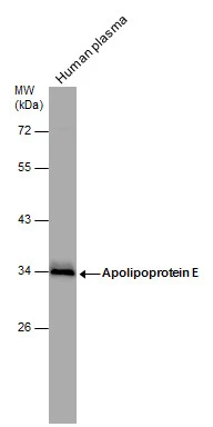
Human tissue extract (30 μg) was separated by 10% SDS-PAGE, and the membrane was blotted with Apolipoprotein E antibody (GTX101456) diluted at 1:1000.
 was separated by 10% SDS-PAGE, and the membrane was blotted with Apolipoprotein E antibody (GTX101456) diluted at 1:1000.")
 dilution: 1:500.
Antigen Retrieval: Trilogy? (EDTA based, pH 8.0) buffer, 15min")
 diluted at 1:500. Blue: Hoechst 33342 staining.")
Human tissue extract (30 μg) was separated by 10% SDS-PAGE, and the membrane was blotted with Apolipoprotein E antibody (GTX101456) diluted at 1:1000.
Apolipoprotein E antibody
GTX101456
ApplicationsImmunoFluorescence, Western Blot, ImmunoCytoChemistry, ImmunoHistoChemistry, ImmunoHistoChemistry Paraffin
Product group Antibodies
ReactivityHuman
TargetAPOE
Overview
- SupplierGeneTex
- Product NameApolipoprotein E antibody
- Delivery Days Customer9
- Application Supplier NoteWB: 1:500-1:3000. ICC/IF: 1:100-1:1000. IHC-P: 1:100-1:1000. *Optimal dilutions/concentrations should be determined by the researcher.Not tested in other applications.
- ApplicationsImmunoFluorescence, Western Blot, ImmunoCytoChemistry, ImmunoHistoChemistry, ImmunoHistoChemistry Paraffin
- CertificationResearch Use Only
- ClonalityPolyclonal
- Concentration1 mg/ml
- ConjugateUnconjugated
- Gene ID348
- Target nameAPOE
- Target descriptionapolipoprotein E
- Target synonymsAD2, APO-E, ApoE4, LDLCQ5, LPG, apolipoprotein E, apolipoprotein E3
- HostRabbit
- IsotypeIgG
- Protein IDP02649
- Protein NameApolipoprotein E
- Scientific DescriptionChylomicron remnants and very low density lipoprotein (VLDL) remnants are rapidly removed from the circulation by receptor-mediated endocytosis in the liver. Apolipoprotein E, a main apoprotein of the chylomicron, binds to a specific receptor on liver cells and peripheral cells. ApoE is essential for the normal catabolism of triglyceride-rich lipoprotein constituents. The APOE gene is mapped to chromosome 19 in a cluster with APOC1 and APOC2. Defects in apolipoprotein E result in familial dysbetalipoproteinemia, or type III hyperlipoproteinemia (HLP III), in which increased plasma cholesterol and triglycerides are the consequence of impaired clearance of chylomicron and VLDL remnants. [provided by RefSeq]
- ReactivityHuman
- Storage Instruction-20°C or -80°C,2°C to 8°C
- UNSPSC12352203
Datasheet
Related products
Product group Antibodies

Anti-Apo E [1D7]Ab01194-1.1
ApplicationsELISA, Other Application, RadioImmunoAssay
ReactivityHuman
TargetAPOE
- SizePrice
Product group Antibodies
Anti-APOE (233-252aa) Antibody130-10506
ApplicationsELISA
ReactivityHuman
TargetAPOE
- SizePrice

Product group Antibodies
Anti-APOE Antibody Picoband(r)A00015-5-CARRIER-FREE
ApplicationsFlow Cytometry, ImmunoFluorescence, Western Blot, ELISA, ImmunoCytoChemistry
ReactivityHuman
TargetAPOE
- SizePrice

Product group Antibodies
ApplicationsWestern Blot, ELISA, ELISpot Assay
ReactivityHuman
TargetAPOE
- SizePrice
![Sandwich ELISA analysis of monkey apoE protein using GTX02906 Apolipoprotein E antibody [E981] as coating antibody and GTX02905-02 Apolipoprotein E antibody [E887] (Biotin) as detecting antibody.](https://www.genetex.com/upload/website/prouct_img/normal/GTX02905-02/GTX02905-02_20210507_ELISA_4_w_23053123_138.webp)
Product group Antibodies
Apolipoprotein E antibody [E887] (Biotin)GTX02905-02
ApplicationsImmunoPrecipitation, Western Blot, ELISA
ReactivityHuman, Monkey
TargetAPOE
- SizePrice
![Sandwich ELISA analysis of monkey apoE protein using GTX02906 Apolipoprotein E antibody [E981] as coating antibody and GTX02905-02 Apolipoprotein E antibody [E887] (Biotin) as detecting antibody.](https://www.genetex.com/upload/website/prouct_img/normal/GTX02906/GTX02906_20210507_ELISA_w_23053123_676.webp)
Product group Antibodies
Apolipoprotein E antibody [E981]GTX02906
ApplicationsWestern Blot, ELISA
ReactivityHuman, Monkey
TargetAPOE
- SizePrice
![Sandwich ELISA analysis of monkey apoE protein using GTX02906 Apolipoprotein E antibody [E981] as coating antibody and GTX02905-02 Apolipoprotein E antibody [E887] (Biotin) as detecting antibody.](https://www.genetex.com/upload/website/prouct_img/normal/GTX03051/GTX03051_20210507_ELISA_w_23053123_346.webp)
Product group Antibodies
ApplicationsELISA
ReactivityHuman, Monkey
TargetAPOE
- SizePrice
![Apolipoprotein E antibody [C2C3], C-term detects Apolipoprotein E protein at cytoplasm by immunofluorescent analysis. Sample: HepG2 cells were fixed in 4% paraformaldehyde at RT for 15 min. Green: Apolipoprotein E stained by Apolipoprotein E antibody [C2C3], C-term (GTX100053) diluted at 1:500. Blue: Hoechst 33342 staining.](https://www.genetex.com/upload/website/prouct_img/normal/GTX100053/GTX100053_43006_20171220_ICC_IF_w_23053123_454.webp)
Product group Antibodies
References
ApplicationsImmunoFluorescence, ImmunoPrecipitation, Western Blot, ELISA, ImmunoCytoChemistry, ImmunoHistoChemistry, ImmunoHistoChemistry Frozen, ImmunoHistoChemistry Paraffin
ReactivityHuman
TargetAPOE
- SizePrice
![FACS analysis of HepG2 cells using GTX60446 Apolipoprotein E antibody [1H4]. Green : Apolipoprotein E Purple : negative control](https://www.genetex.com/upload/website/prouct_img/normal/GTX60446/GTX60446_20170912_FACS_w_23061123_124.webp)
Product group Antibodies
References
Apolipoprotein E antibody [1H4]GTX60446
ApplicationsFlow Cytometry, Western Blot, ELISA, ImmunoHistoChemistry, ImmunoHistoChemistry Paraffin
ReactivityHuman
TargetAPOE
- SizePrice
![Human plasma (30 μg) was separated by 10% SDS-PAGE, and the membrane was blotted with Apolipoprotein E antibody [GT27711] (GTX635889) diluted at 1:25000. The HRP-conjugated anti-mouse IgG antibody (GTX213111-01) was used to detect the primary antibody.](https://www.genetex.com/upload/website/prouct_img/normal/GTX635889/GTX635889_44195_20220225_WB_plasma_w_23061202_472.webp)
Product group Antibodies
Apolipoprotein E antibody [GT27711]GTX635889
ApplicationsImmunoPrecipitation, Western Blot, ELISA
ReactivityHuman, Rat
TargetAPOE
- SizePrice