

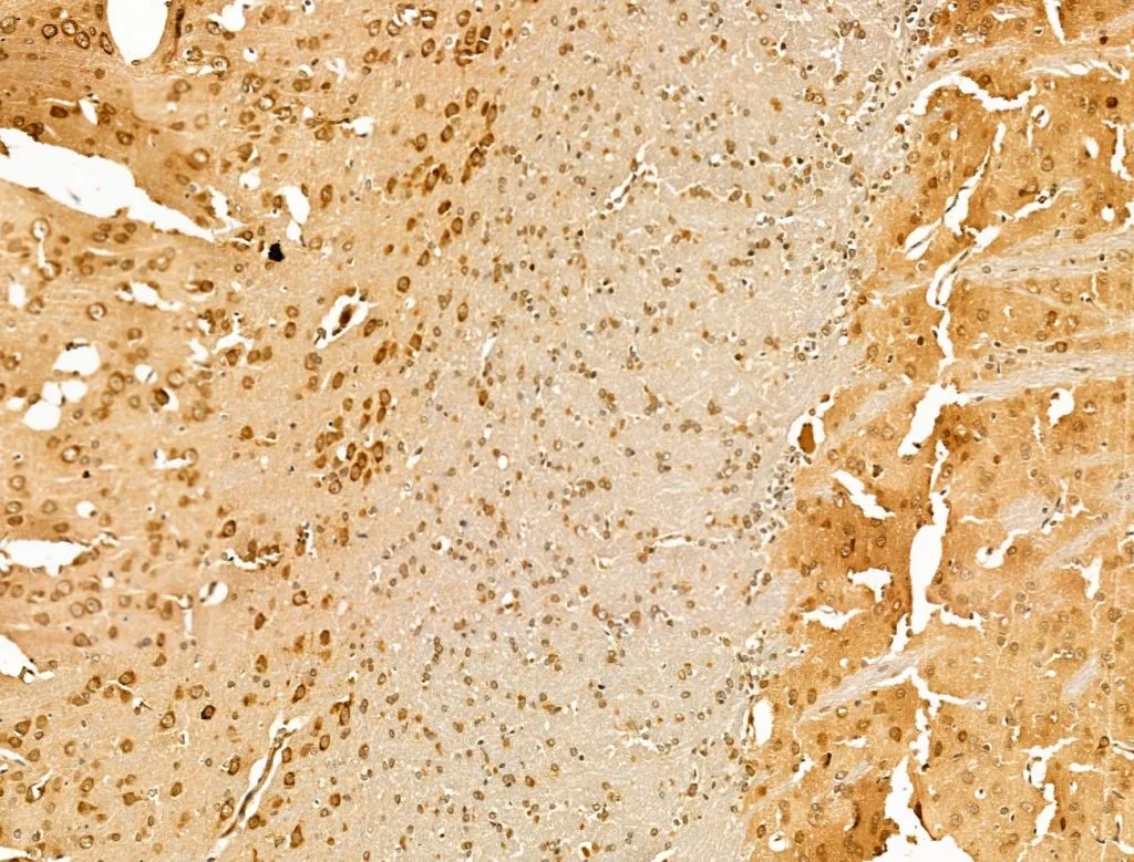
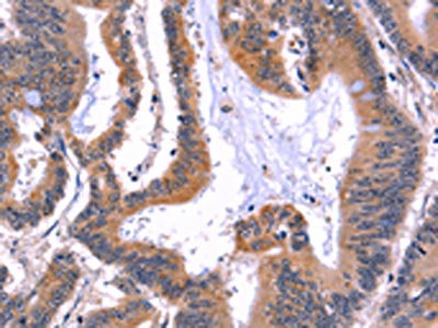
IHC-P analysis of rat brain tissue using GTX04096 ARC antibody. Antigen retrieval : Heat mediated antigen retrieval step in citrate buffer Dilution : 1:100


IHC-P analysis of rat brain tissue using GTX04096 ARC antibody. Antigen retrieval : Heat mediated antigen retrieval step in citrate buffer Dilution : 1:100
ARC antibody
GTX04096
ApplicationsImmunoFluorescence, Western Blot, ImmunoCytoChemistry, ImmunoHistoChemistry, ImmunoHistoChemistry Paraffin
Product group Antibodies
ReactivityHuman, Mouse, Rat
TargetARC
Overview
- SupplierGeneTex
- Product NameARC antibody
- Delivery Days Customer9
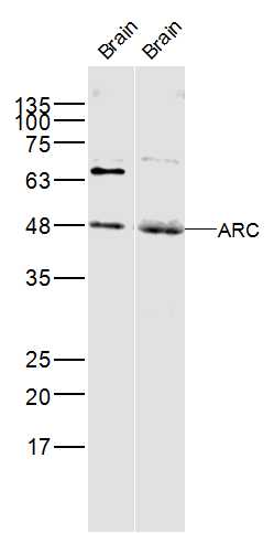
- Application Supplier NoteWB: 1:500-1:2000. ICC/IF: 1:100-1:500. IHC-P: 1:50-1:200. *Optimal dilutions/concentrations should be determined by the researcher.Not tested in other applications.
- ApplicationsImmunoFluorescence, Western Blot, ImmunoCytoChemistry, ImmunoHistoChemistry, ImmunoHistoChemistry Paraffin
- CertificationResearch Use Only
- ClonalityPolyclonal
- Concentration1 mg/ml
- ConjugateUnconjugated
- Gene ID23237
- Target nameARC
- Target descriptionactivity regulated cytoskeleton associated protein
- Target synonymsArg3.1, hArc, activity-regulated cytoskeleton-associated protein, ARC/ARG3.1, activity-regulated gene 3.1 protein homolog
- HostRabbit
- IsotypeIgG
- Protein IDQ7LC44
- Protein NameActivity-regulated cytoskeleton-associated protein
- Scientific DescriptionPredicted to enable mRNA binding activity. Involved in cell migration; cytoskeleton organization; and regulation of cell morphogenesis. Located in cytoplasm and plasma membrane. [provided by Alliance of Genome Resources, Apr 2022]
- ReactivityHuman, Mouse, Rat
- Storage Instruction-20°C or -80°C,2°C to 8°C
- UNSPSC41116161
Datasheet
Related products

Product group Antibodies
Anti-Arc AntibodyA10637
ApplicationsWestern Blot
ReactivityHuman, Mouse
- SizePrice
Product group Antibodies
Anti-ARC Antibody144-66726
ApplicationsWestern Blot
ReactivityHuman, Mouse
TargetARC
- SizePrice
Product group Antibodies
References
ARC Polyclonal AntibodyBS-0385R
ApplicationsFlow Cytometry, Western Blot, ELISA
ReactivityBovine, Equine, Human, Mouse, Rat
TargetARC
- SizePrice
Product group Antibodies
ARC AntibodyCSB-PA698018
ApplicationsWestern Blot, ELISA, ImmunoHistoChemistry
ReactivityHuman, Mouse, Rat
TargetARC
- SizePrice
Product group Antibodies
ApplicationsImmunoPrecipitation, Western Blot, ImmunoCytoChemistry, ImmunoHistoChemistry
TargetARC
- SizePrice
Product group Antibodies

ARC / Arg3.1 AntibodyLS-C401272
ApplicationsELISA, ImmunoHistoChemistry
ReactivityHuman
TargetARC
- SizePrice

Product group Antibodies
Anti-ARC AntibodyHPA056430
ApplicationsImmunoCytoChemistry
ReactivityHuman
TargetARC
- SizePrice
Product group Antibodies
Anti-ARCY058421
ApplicationsWestern Blot, ImmunoHistoChemistry
ReactivityHuman, Mouse
- SizePrice
Product group Antibodies
Anti-ARC AntibodyCAB9177
ApplicationsWestern Blot, ELISA
ReactivityHuman
TargetARC
- SizePrice

Product group Antibodies
Anti-Arc Antibody Picoband(r)PB9753-CARRIER-FREE
ApplicationsWestern Blot
ReactivityHuman, Mouse, Rat
TargetARC
- SizePrice