Bio-Connect
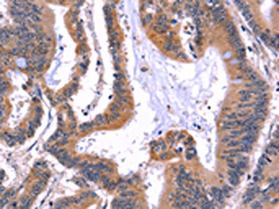
Immunohistochemistry of paraffin-embedded Human colon cancer tissue using ARC Polyclonal Antibody at dilution 1:50

ARC Polyclonal Antibody

E-AB-10826
Elabscience
Product group Antibodies
Sign in to order and to see your custom pricing.
Large volume orders?
Order with a bulk request

Overview

  • Supplier
    Elabscience
  • Product Name
    ARC Polyclonal Antibody
  • Delivery Days Customer
    12
  • Applications Supplier
    ELISA IHC
  • Certification
    Research Use Only
  • Concentration
    0.5mg/ml
  • Scientific Description
    Arc (for activity-regulated cytoskeleton-associated protein) is a growth factor and immediate early gene that is enriched in brain. Arc mRNA and protein levels are induced by neuronal activity, which is necessary to stimulate neuroplasticity, indicating a potential role for Arc in activity-dependent changes in dendrite function. Arc expression has been detected in neuronal cell bodies and dendrites in the hippocampus, amygdala, hypothalamus, striatum and cortex. Arc has been shown to localize to the cytoskeleton of neuronal cells and appears to colocalize with F-Actin, although it may associate with an Actin-associated protein rather than directly with F-Actin. It has been shown that cocaine-stimulated neuronal activity results in increased Arc mRNA levels in striatum.
  • UNSPSC
    12352203

Related products

The image on the left is immunohistochemistry of paraffin-embedded Human colon cancer tissue using CSB-PA698018(ARC Antibody) at dilution 1/50, on the right is treated with fusion protein. (Original magnification: x200)
Product group Antibodies
Cusabio
ARC AntibodyCSB-PA698018
ApplicationsWestern Blot, ELISA, ImmunoHistoChemistry
TargetARC
  • SizePrice
Product group Antibodies
Assay Genie
TargetARC
  • SizePrice
Product group Antibodies
Biomatik
ApplicationsImmunoPrecipitation, Western Blot, ImmunoCytoChemistry, ImmunoHistoChemistry
TargetARC
  • SizePrice
Lane 1: Rat brain; Lane 2: mouse brain lysate probed with ARC Polyclonal Antibody, Unconjugated (bs-0385R) at 1:300 dilution and 4˚C overnight incubation. Followed by conjugated secondary antibody incubation at 1:10000 for 60 min at 37˚C.
Product group Antibodies
Bioss Antibodies
ApplicationsFlow Cytometry, Western Blot, ELISA
TargetARC
  • SizePrice
Product group Antibodies
RayBiotech
ApplicationsWestern Blot
TargetARC
  • SizePrice
Immunofluorescent staining of human cell line U-2 OS shows localization to microtubules & vesicles.
Product group Antibodies
Atlas Antibodies
ApplicationsImmunoCytoChemistry
TargetARC
  • SizePrice
ICC/IF analysis of PFA-fixed HeLa cells using GTX04096 ARC antibody. Red : Primary antibody Green : beta tubulin Blue : DAPI Permeabilize : 0.1% Triton X-100 Dilution : 1:200
Product group Antibodies
GeneTex
ARC antibodyGTX04096
ApplicationsImmunoFluorescence, Western Blot, ImmunoCytoChemistry, ImmunoHistoChemistry, ImmunoHistoChemistry Paraffin
TargetARC
  • SizePrice
Anti-Arc Antibody
Product group Antibodies
Antibodies.com
ApplicationsWestern Blot
  • SizePrice
Product group Antibodies
LifeSpan BioSciences
ApplicationsELISA, ImmunoHistoChemistry
TargetARC
  • SizePrice
Figure 1. Western blot analysis of Arc using anti-Arc antibody (PB9753). Electrophoresis was performed on a 5-20% SDS-PAGE gel at 70V (Stacking gel) / 90V (Resolving gel) for 2-3 hours. The sample well of each lane was loaded with 30 ug of sample under reducing conditions. Lane 1: human U20S whole cell lysates, Lane 2: human 293T whole cell lysates, Lane 3: human SH-SY5Y whole cell lysates, Lane 4: human SiHa whole cell lysates, Lane 5: rat brain tissue lysates, Lane 6: rat C6 whole cell lysates, Lane 7: mouse brain tissue lysates, Lane 8: mouse Neuro-2a whole cell lysates. After electrophoresis, proteins were transferred to a nitrocellulose membrane at 150 mA for 50-90 minutes. Blocked the membrane with 5% non-fat milk/TBS for 1.5 hour at RT. The membrane was incubated with rabbit anti-Arc antigen affinity purified polyclonal antibody (Catalog # PB9753) at 0.5 microg/mL overnight at 4°C, then washed with TBS-0.1%Tween 3 times with 5 minutes each and probed with a goat anti-rabbit IgG-HRP secondary antibody at a dilution of 1:5000 for 1.5 hour at RT. The signal is developed using an Enhanced Chemiluminescent detection (ECL) kit (Catalog # EK1002) with Tanon 5200 system. A specific band was detected for Arc at approximately 45 kDa. The expected band size for Arc is at 45 kDa.
Product group Antibodies
Boster Bio
ApplicationsWestern Blot
TargetARC
  • SizePrice