

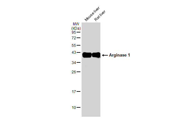
Various tissue extracts (50 μg) were separated by 12% SDS-PAGE, and the membrane was blotted with Arginase 1 antibody (GTX109242) diluted at 1:1000. The HRP-conjugated anti-rabbit IgG antibody (GTX213110-01) was used to detect the primary antibody.
 (GTX213110-01) was diluted at 1:10000 and used to detect the primary antibody.")
 diluted at 1:500. Antigen Retrieval: Citrate buffer, pH 6.0, 15 min")
. Western blot analysis was performed using Arginase 1 antibody (GTX109242) diluted at 1:600. EasyBlot anti-Rabbit IgG (GTX221666-01) was used as a secondary reagent.")
 detects ARG1 protein by flow cytometry analysis. Sample: HepG2 cell. Black: Unlabelled sample was used as a control. Red: Arginase 1 antibody (GTX109242) dilution: 1:50. Acquisition of 20,000 events were collected for FACS analysis.")
 (GTX213111-01) was diluted at 1:10000 and used to detect the primary antibody.")
 diluted at 1:500. Blue: Fluoroshield with DAPI (GTX30920). Scale bar= 10 μm.")
 and transfected (+) HepG2 whole cell extracts (30 μg) were separated by 12% SDS-PAGE, and the membrane was blotted with Arginase 1 antibody (GTX109242) diluted at 1:500. The HRP-conjugated anti-rabbit IgG antibody (GTX213110-01) was used to detect the primary antibody.")


Various tissue extracts (50 μg) were separated by 12% SDS-PAGE, and the membrane was blotted with Arginase 1 antibody (GTX109242) diluted at 1:1000. The HRP-conjugated anti-rabbit IgG antibody (GTX213110-01) was used to detect the primary antibody.
Arginase 1 antibody
GTX109242
ApplicationsFlow Cytometry, ImmunoFluorescence, ImmunoPrecipitation, Western Blot, ELISA, ImmunoCytoChemistry, ImmunoHistoChemistry, ImmunoHistoChemistry Frozen, ImmunoHistoChemistry Paraffin
Product group Antibodies
ReactivityHuman, Mouse, Rat
TargetARG1
Overview
- SupplierGeneTex
- Product NameArginase 1 antibody
- Delivery Days Customer9
- Application Supplier NoteWB: 1:500-1:3000. ICC/IF: 1:100-1:1000. IHC-P: 1:100-1:1000. IHC-Fr: 1:100-1:1000. FACS: 1:50-1:200. IP: 1:100-1:1000. ELISA: 1:1000-1:10000. *Optimal dilutions/concentrations should be determined by the researcher.Not tested in other applications.
- ApplicationsFlow Cytometry, ImmunoFluorescence, ImmunoPrecipitation, Western Blot, ELISA, ImmunoCytoChemistry, ImmunoHistoChemistry, ImmunoHistoChemistry Frozen, ImmunoHistoChemistry Paraffin
- CertificationResearch Use Only
- ClonalityPolyclonal
- Concentration0.72 mg/ml
- ConjugateUnconjugated
- Gene ID383
- Target nameARG1
- Target descriptionarginase 1
- Target synonymsarginase-1, arginase, liver, liver-type arginase, type I arginase
- HostRabbit
- IsotypeIgG
- Protein IDP05089
- Protein NameArginase-1
- Scientific DescriptionArginase catalyzes the hydrolysis of arginine to ornithine and urea. At least two isoforms of mammalian arginase exist (types I and II) which differ in their tissue distribution, subcellular localization, immunologic crossreactivity and physiologic function. The type I isoform encoded by this gene, is a cytosolic enzyme and expressed predominantly in the liver as a component of the urea cycle. Inherited deficiency of this enzyme results in argininemia, an autosomal recessive disorder characterized by hyperammonemia. [provided by RefSeq]
- ReactivityHuman, Mouse, Rat
- Storage Instruction-20°C or -80°C,2°C to 8°C
- UNSPSC12352203
References
- Hofmann L, Harasymczuk M, Huber D, et al. Arginase-1 in Plasma-Derived Exosomes as Marker of Metastasis in Patients with Head and Neck Squamous Cell Carcinoma. Cancers (Basel). 2023,15(22). doi: 10.3390/cancers15225449Read this paper
- Bokil AA, Le Boulvais Børkja M, Wolowczyk C, et al. Discovery of a new marker to identify myeloid cells associated with metastatic breast tumours. Cancer Cell Int. 2023,23(1):279. doi: 10.1186/s12935-023-03136-wRead this paper
- Mossmann D, Müller C, Park S, et al. Arginine reprograms metabolism in liver cancer via RBM39. Cell. 2023,186(23):5068-5083.e23. doi: 10.1016/j.cell.2023.09.011Read this paper
- Shosha E, Shahror RA, Morris CA, et al. The arginase 1/ornithine decarboxylase pathway suppresses HDAC3 to ameliorate the myeloid cell inflammatory response: implications for retinal ischemic injury. Cell Death Dis. 2023,14(9):621. doi: 10.1038/s41419-023-06147-7Read this paper
- Yang JX, Zhu J, Ni K, et al. Electroacupuncture relieves chronic pain by promoting microglia M2 polarization in lumbar disc herniation rats. Neuroreport. 2023,34(12):638-648. doi: 10.1097/WNR.0000000000001935Read this paper
- Panebianco C, Pisati F, Villani A, et al. Counteracting gemcitabine+nab-paclitaxel induced dysbiosis in KRAS wild type and KRAS(G12D) mutated pancreatic cancer in vivo model. Cell Death Discov. 2023,9(1):116. doi: 10.1038/s41420-023-01397-yRead this paper
- Huang J, Tao H, Yancey PG, et al. Scavenging dicarbonyls with 5'-O-pentyl-pyridoxamine increases HDL net cholesterol efflux capacity and attenuates atherosclerosis and insulin resistance. Mol Metab. 2023,67:101651. doi: 10.1016/j.molmet.2022.101651Read this paper
- Schaafsma E, Croteau W, ElTanbouly M, et al. VISTA Targeting of T-cell Quiescence and Myeloid Suppression Overcomes Adaptive Resistance. Cancer Immunol Res. 2023,11(1):38-55. doi: 10.1158/2326-6066.CIR-22-0116Read this paper
- Anderson S, Prateeksha P, Das H. Dental Pulp-Derived Stem Cells Reduce Inflammation, Accelerate Wound Healing and Mediate M2 Polarization of Myeloid Cells. Biomedicines. 2022,10(8). doi: 10.3390/biomedicines10081999Read this paper
- Liu L, Fang L, Duan B, et al. Multi-Hit White Matter Injury-Induced Cerebral Palsy Model Established by Perinatal Lipopolysaccharide Injection. Front Pediatr. 2022,10:867410. doi: 10.3389/fped.2022.867410Read this paper
Datasheet
Related products
Product group Antibodies

Anti-Arginase 1 [mAb5]Ab03081-1.1
ApplicationsElectron Microscopy, Neutralisation/Blocking
ReactivityHuman
TargetARG1
- SizePrice

Product group Antibodies
Anti-ARG1 Antibody144-01847
ApplicationsWestern Blot, ImmunoHistoChemistry
ReactivityHuman, Mouse, Rat
TargetARG1
- SizePrice

Product group Antibodies
Anti-ARG1 AntibodyAMAB90545
ApplicationsWestern Blot, ImmunoHistoChemistry
ReactivityHuman
TargetARG1
- SizePrice

Product group Antibodies
Anti-liver Arginase/ARG1 Antibody Picoband(r)A01106-CARRIER-FREE
ApplicationsWestern Blot, ELISA, ImmunoHistoChemistry
ReactivityHuman, Monkey, Mouse, Rat
TargetARG1
- SizePrice
![IHC-P analysis of human liver tissue using GTX04426 Arginase 1 antibody [MSVA-511R] HistoMAX?. Strong nuclear and cytoplasmic arginase 1 expression in all hepatocytes in a normal liver.](https://www.genetex.com/upload/website/prouct_img/normal/GTX04426/GTX04426_20230728_IHC-P_4_23072722_512.webp)
Product group Antibodies
ApplicationsImmunoHistoChemistry, ImmunoHistoChemistry Paraffin
ReactivityHuman
TargetARG1
- SizePrice
Product group Antibodies
References
Arginase 1 antibodyGTX113131
ApplicationsImmunoFluorescence, Western Blot, ELISA, ImmunoCytoChemistry, ImmunoHistoChemistry, ImmunoHistoChemistry Paraffin
ReactivityHuman, Mouse, Rat
TargetARG1
- SizePrice
Product group Antibodies
References
Arginase 1 antibody [GT5811]GTX634218
ApplicationsFlow Cytometry, Western Blot, ELISA, ImmunoHistoChemistry, ImmunoHistoChemistry Paraffin
ReactivityHuman, Mouse, Rat
TargetARG1
- SizePrice
![Non-transfected (–) and transfected (+) 293T whole cell extracts (30 μg) were separated by 12% SDS-PAGE, and the membrane was blotted with Arginase 1 antibody [HL1891] (GTX637640) diluted at 1:2500. The HRP-conjugated anti-rabbit IgG antibody (GTX213110-01) was used to detect the primary antibody.](https://www.genetex.com/upload/website/prouct_img/normal/GTX637640/GTX637640_T-44837_20230203_WB_multiple_B_23020621_412.webp)
Product group Antibodies
Arginase 1 antibody [HL1891]GTX637640
ApplicationsWestern Blot, ImmunoHistoChemistry, ImmunoHistoChemistry Paraffin
ReactivityHuman, Mouse, Rat
TargetARG1
- SizePrice
Product group Antibodies
ARG1 Polyclonal AntibodyCAC14814
ApplicationsWestern Blot, ELISA, ImmunoHistoChemistry
ReactivityMouse, Rat
TargetARG1
- SizePrice