

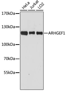
WB analysis of various sample lysates using GTX35236 ARHGEF1 antibody. Dilution : 1:3000 Loading : 25μg per lane

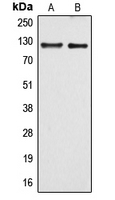
 and knockout (KO) 293T cell lysate using GTX35236 ARHGEF1 antibody. Dilution : 1:500 Loading : 25μg per lane")
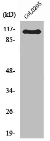
WB analysis of various sample lysates using GTX35236 ARHGEF1 antibody. Dilution : 1:3000 Loading : 25μg per lane
ARHGEF1 antibody
GTX35236
ApplicationsWestern Blot, ImmunoHistoChemistry, ImmunoHistoChemistry Paraffin
Product group Antibodies
ReactivityHuman
TargetARHGEF1
Overview
- SupplierGeneTex
- Product NameARHGEF1 antibody
- Delivery Days Customer9
- Application Supplier NoteWB: 1:500 - 1:2000. IHC-P: 1:50 - 1:100. *Optimal dilutions/concentrations should be determined by the researcher.Not tested in other applications.
- ApplicationsWestern Blot, ImmunoHistoChemistry, ImmunoHistoChemistry Paraffin
- CertificationResearch Use Only
- ClonalityPolyclonal
- ConjugateUnconjugated
- Gene ID9138
- Target nameARHGEF1
- Target descriptionRho guanine nucleotide exchange factor 1
- Target synonymsGEF1, IMD62, LBCL2, LSC, P115-RHOGEF, SUB1.5, rho guanine nucleotide exchange factor 1, 115 kDa guanine nucleotide exchange factor, 115-kD protein, Lsc homolog, Rho guanine nucleotide exchange factor (GEF) 1, p115RhoGEF
- HostRabbit
- IsotypeIgG
- Protein IDQ92888
- Protein NameRho guanine nucleotide exchange factor 1
- Scientific DescriptionRho GTPases play a fundamental role in numerous cellular processes that are initiated by extracellular stimuli that work through G protein coupled receptors. The encoded protein may form complex with G proteins and stimulate Rho-dependent signals. Multiple alternatively spliced transcript variants have been found for this gene, but the full-length nature of some variants has not been defined. [provided by RefSeq, Jul 2008]
- ReactivityHuman
- Storage Instruction-20°C or -80°C,2°C to 8°C
- UNSPSC41116161
Datasheet
Related products

Product group Antibodies
Anti-ARHGEF1 AntibodyA96598
ApplicationsWestern Blot, ELISA
ReactivityHuman, Mouse, Rat
- SizePrice
Product group Antibodies
Anti-ARHGEF1 Antibody144-61386
ApplicationsWestern Blot, ImmunoHistoChemistry
ReactivityHuman
TargetARHGEF1
- SizePrice
Product group Antibodies
ARHGEF1 Polyclonal AntibodyBS-55014R
ApplicationsImmunoFluorescence, Western Blot, ImmunoHistoChemistry, ImmunoHistoChemistry Paraffin
ReactivityHuman, Rat
TargetARHGEF1
- SizePrice
Product group Antibodies
ARHGEF1 AntibodyCSB-PA003977
ApplicationsWestern Blot, ELISA
ReactivityHuman, Mouse, Rat
TargetARHGEF1
- SizePrice
Product group Antibodies

ARHGEF1 AntibodyLS-C401278
ApplicationsELISA, ImmunoHistoChemistry
ReactivityHuman, Mouse, Rat
TargetARHGEF1
- SizePrice
Product group Antibodies
ARHGEF1 antibodyGTX55910
ApplicationsWestern Blot
ReactivityHuman, Mouse
TargetARHGEF1
- SizePrice

Product group Antibodies
Anti-ARHGEF1 AntibodyHPA012924
ApplicationsWestern Blot, ImmunoCytoChemistry, ImmunoHistoChemistry
ReactivityHuman
TargetARHGEF1
- SizePrice
Product group Antibodies
ApplicationsWestern Blot, ELISA, ImmunoHistoChemistry, ImmunoHistoChemistry Paraffin
ReactivityHuman
TargetARHGEF1
- SizePrice

Product group Antibodies
Anti-ARHGEF1 Antibody Picoband(r)PB10045-CARRIER-FREE
ApplicationsFlow Cytometry, ImmunoFluorescence, Western Blot, ImmunoCytoChemistry, ImmunoHistoChemistry
ReactivityHamster, Human, Mouse, Rat
TargetARHGEF1
- SizePrice