

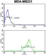
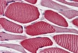
FACS analysis of MDA-MB231 cells using GTX81198 AS160 / TBC1D4 antibody, N-term. Top histogram : negative control Bottom histogram : MDA-MB231 cells
 using GTX81198 AS160 / TBC1D4 antibody, N-term.")

FACS analysis of MDA-MB231 cells using GTX81198 AS160 / TBC1D4 antibody, N-term. Top histogram : negative control Bottom histogram : MDA-MB231 cells
AS160 / TBC1D4 antibody, N-term
GTX81198
ApplicationsFlow Cytometry, Western Blot, ImmunoHistoChemistry, ImmunoHistoChemistry Paraffin
Product group Antibodies
ReactivityHuman
TargetTBC1D4
Overview
- SupplierGeneTex
- Product NameAS160 / TBC1D4 antibody, N-term
- Delivery Days Customer9
- Application Supplier NoteWB: 1:1000. IHC-P: 1:50-1:100. FCM: 1:10-1:50. *Optimal dilutions/concentrations should be determined by the researcher.Not tested in other applications.
- ApplicationsFlow Cytometry, Western Blot, ImmunoHistoChemistry, ImmunoHistoChemistry Paraffin
- CertificationResearch Use Only
- ClonalityPolyclonal
- ConjugateUnconjugated
- Gene ID9882
- Target nameTBC1D4
- Target descriptionTBC1 domain family member 4
- Target synonymsAS160, NIDDM5, TBC1 domain family member 4, TBC (Tre-2, BUB2, CDC16) domain-containing protein, akt substrate of 160 kDa
- HostRabbit
- IsotypeIgG
- Protein IDO60343
- Protein NameTBC1 domain family member 4
- Scientific DescriptionThis gene is a member of the Tre-2/BUB2/CDC16 domain family. The protein encoded by this gene is a Rab-GTPase-activating protein, and contains two phopshotyrosine-binding domains (PTB1 and PTB2), a calmodulin-binding domain (CBD), a Rab-GTPase domain, and multiple AKT phosphomotifs. This protein is thought to play an important role in glucose homeostasis by regulating the insulin-dependent trafficking of the glucose transporter 4 (GLUT4), important for removing glucose from the bloodstream into skeletal muscle and fat tissues. Reduced expression of this gene results in an increase in GLUT4 levels at the plasma membrane, suggesting that this protein is important in intracellular retention of GLUT4 under basal conditions. When exposed to insulin, this protein is phosphorylated, dissociates from GLUT4 vesicles, resulting in increased GLUT4 at the cell surface, and enhanced glucose transport. Phosphorylation of this protein by AKT is required for proper translocation of GLUT4 to the cell surface. Individuals homozygous for a mutation in this gene are at higher risk for type 2 diabetes and have higher levels of circulating glucose and insulin levels after glucose ingestion. Alternative splicing results in multiple transcript variants encoding different isoforms. [provided by RefSeq, Aug 2015]
- ReactivityHuman
- Storage Instruction-20°C or -80°C,2°C to 8°C
- UNSPSC41116161
Datasheet
Related products
Product group Antibodies
Anti-AS160 AntibodyA99172
ApplicationsImmunoFluorescence, ELISA
ReactivityHuman, Mouse
- SizePrice
Product group Antibodies
Anti-TBC1D4 Antibody144-65316
ApplicationsImmunoFluorescence, Western Blot
ReactivityHuman, Mouse, Rat
TargetTBC1D4
- SizePrice

Product group Antibodies
Anti-AS160/TBC1D4 Antibody Picoband(r)A02004-3-CARRIER-FREE
ApplicationsFlow Cytometry, ImmunoFluorescence, Western Blot, ELISA, ImmunoCytoChemistry, ImmunoHistoChemistry
ReactivityHuman
TargetTBC1D4
- SizePrice
Product group Antibodies
TBC1D4 Polyclonal AntibodyBS-5893R
ApplicationsImmunoFluorescence, ELISA, ImmunoCytoChemistry, ImmunoHistoChemistry, ImmunoHistoChemistry Frozen, ImmunoHistoChemistry Paraffin
ReactivityBovine, Human, Mouse, Porcine, Rat, Sheep
TargetTBC1D4
- SizePrice
Product group Antibodies

Goat anti-AS160 / TBC1D4EB07591
ApplicationsWestern Blot, ELISA, ImmunoHistoChemistry
ReactivityCanine, Human
TargetTBC1D4
- SizePrice
Product group Antibodies
TBC1D4 AntibodyCSB-PA023218LA01HU
ApplicationsImmunoFluorescence, ELISA, ImmunoHistoChemistry
ReactivityHuman
TargetTBC1D4
- SizePrice
Product group Antibodies

TBC1D4 / AS160 AntibodyLS-C403640
ApplicationsWestern Blot, ELISA, ImmunoHistoChemistry
ReactivityHuman, Mouse
TargetTBC1D4
- SizePrice
Product group Antibodies
AS160 / TBC1D4 antibodyGTX31767
ApplicationsELISA, ImmunoHistoChemistry, ImmunoHistoChemistry Paraffin
ReactivityHuman
TargetTBC1D4
- SizePrice
Product group Antibodies
ApplicationsWestern Blot, ImmunoHistoChemistry, ImmunoHistoChemistry Paraffin
ReactivityHuman
TargetTBC1D4
- SizePrice