

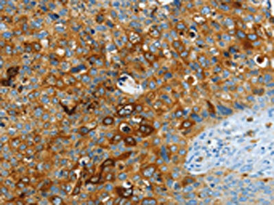
The image on the left is immunohistochemistry of paraffin-embedded Human cervical cancer tissue using CSB-PA208943(ASAH1 Antibody) at dilution 1/40, on the right is treated with fusion protein. (Original magnification: x200)
 at dilution 1/40, on the right is treated with fusion protein. (Original magnification: x200)")
 at dilution 1/800, Secondary antibody: Goat anti rabbit IgG at 1/8000 dilution, Exposure time: 30 seconds")
The image on the left is immunohistochemistry of paraffin-embedded Human cervical cancer tissue using CSB-PA208943(ASAH1 Antibody) at dilution 1/40, on the right is treated with fusion protein. (Original magnification: x200)
ASAH1 Antibody
CSB-PA208943
ApplicationsWestern Blot, ELISA, ImmunoHistoChemistry
Product group Antibodies
ReactivityHuman, Mouse, Rat
TargetASAH1
Overview
- SupplierCusabio
- Product NameASAH1 Antibody
- Delivery Days Customer20
- ApplicationsWestern Blot, ELISA, ImmunoHistoChemistry
- CertificationResearch Use Only
- ClonalityPolyclonal
- ConjugateUnconjugated
- Gene ID427
- Target nameASAH1
- Target descriptionN-acylsphingosine amidohydrolase 1
- Target synonymsAC, ACDase, ASAH, PHP, PHP32, SMAPME, acid ceramidase, N-acylethanolamine hydrolase ASAH1, N-acylsphingosine amidohydrolase (acid ceramidase) 1, acid CDase, acylsphingosine deacylase, glycosylceramide deacylase, putative 32 kDa heart protein
- HostRabbit
- IsotypeIgG
- Protein IDQ13510
- Protein NameAcid ceramidase
- Scientific DescriptionThis gene encodes a heterodimeric protein consisting of a nonglycosylated alpha subunit and a glycosylated beta subunit that is cleaved to the mature enzyme posttranslationally. The encoded protein catalyzes the synthesis and degradation of ceramide into sphingosine and fatty acid. Mutations in this gene have been associated with a lysosomal storage disorder known as Farber disease. Multiple transcript variants encoding several distinct isoforms have been identified for this gene.
- ReactivityHuman, Mouse, Rat
- Storage Instruction-20°C or -80°C
- UNSPSC41116161
Related products
Product group Antibodies
Anti-ASAH1 AntibodyA9903
ApplicationsWestern Blot
ReactivityMouse, Rat
- SizePrice
Product group Antibodies
Anti-ASAH1 Antibody144-63003
ApplicationsWestern Blot
ReactivityHuman, Mouse, Rat
TargetASAH1
- SizePrice

Product group Antibodies
Anti-ASAH1 Picoband(r) AntibodyA02055-1-CARRIER-FREE
ApplicationsImmunoFluorescence, Western Blot, ELISA, ImmunoCytoChemistry, ImmunoHistoChemistry
ReactivityHuman, Mouse, Rat
TargetASAH1
- SizePrice
Product group Antibodies
ASAH1 Polyclonal AntibodyBS-12976R
ApplicationsImmunoFluorescence, Western Blot, ELISA, ImmunoCytoChemistry, ImmunoHistoChemistry, ImmunoHistoChemistry Frozen, ImmunoHistoChemistry Paraffin
ReactivityBovine, Canine, Chicken, Equine, Human, Mouse, Porcine, Rat, Sheep
TargetASAH1
- SizePrice
Product group Antibodies
ASAH1 Polyclonal AntibodyCAC14896
ApplicationsWestern Blot, ELISA, ImmunoHistoChemistry
TargetASAH1
- SizePrice

Product group Antibodies
Anti-ASAH1-25ulHPA005468
ApplicationsWestern Blot, ImmunoHistoChemistry
ReactivityHuman
- SizePrice
Product group Antibodies
ASAH1 antibodyGTX114267
ApplicationsWestern Blot, ImmunoHistoChemistry, ImmunoHistoChemistry Paraffin
ReactivityHuman, Mouse
TargetASAH1
- SizePrice
Product group Antibodies
Anti-ASAH1 AntibodyCAB13948
ApplicationsImmunoFluorescence, Western Blot, ELISA, ImmunoCytoChemistry
ReactivityHuman
TargetASAH1
- SizePrice
Product group Antibodies

ASAH1 / Acid Ceramidase AntibodyLS-C401406
ApplicationsWestern Blot, ELISA, ImmunoHistoChemistry
ReactivityHuman, Mouse, Rat
TargetASAH1
- SizePrice