

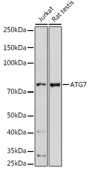
WB analysis of various sample lysates using GTX32459 ATG7 antibody. Dilution : 1:1000 Loading : 25μg per lane


WB analysis of various sample lysates using GTX32459 ATG7 antibody. Dilution : 1:1000 Loading : 25μg per lane
ATG7 antibody
GTX32459
ApplicationsImmunoFluorescence, Western Blot, ImmunoCytoChemistry, ImmunoHistoChemistry, ImmunoHistoChemistry Paraffin
Product group Antibodies
ReactivityHuman, Mouse, Rat
TargetATG7
Overview
- SupplierGeneTex
- Product NameATG7 antibody
- Delivery Days Customer9
- Application Supplier NoteWB: 1:500 - 1:2000. ICC/IF: 1:50 - 1:200. IHC-P: 1:50 - 1:200. *Optimal dilutions/concentrations should be determined by the researcher.Not tested in other applications.
- ApplicationsImmunoFluorescence, Western Blot, ImmunoCytoChemistry, ImmunoHistoChemistry, ImmunoHistoChemistry Paraffin
- CertificationResearch Use Only
- ClonalityPolyclonal
- ConjugateUnconjugated
- Gene ID10533
- Target nameATG7
- Target descriptionautophagy related 7
- Target synonymsAPG7-LIKE, APG7L, GSA7, SCAR31, ubiquitin-like modifier-activating enzyme ATG7, APG7 autophagy 7-like, ATG12-activating enzyme E1 ATG7, hAGP7, ubiquitin-activating enzyme E1-like protein
- HostRabbit
- IsotypeIgG
- Protein IDO95352
- Protein NameUbiquitin-like modifier-activating enzyme ATG7
- Scientific DescriptionThis gene encodes an E1-like activating enzyme that is essential for autophagy and cytoplasmic to vacuole transport. The encoded protein is also thought to modulate p53-dependent cell cycle pathways during prolonged metabolic stress. It has been associated with multiple functions, including axon membrane trafficking, axonal homeostasis, mitophagy, adipose differentiation, and hematopoietic stem cell maintenance. Alternative splicing results in multiple transcript variants. [provided by RefSeq, Sep 2015]
- ReactivityHuman, Mouse, Rat
- Storage Instruction-20°C or -80°C,2°C to 8°C
- UNSPSC41116161
References
- Does the Autophagy Related Gene 7 (ATG7) Have a Role in Non-Melanoma Skin Cancer? Samaka RM et al., 2020, Clin Cosmet Investig DermatolRead this paper
- Trophoblast-Specific Conditional Atg7 Knockout Mice Develop Gestational Hypertension. Aoki A et al., 2018 Nov, Am J PatholRead this paper
Datasheet
Related products
Product group Antibodies
Atg7 Polyclonal AntibodyCAC08251
ApplicationsImmunoFluorescence, Western Blot, ELISA, ImmunoHistoChemistry
TargetATG7
- SizePrice
Product group Antibodies
References
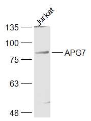
ATG7/APG7 Polyclonal AntibodyBS-2432R
ApplicationsImmunoFluorescence, Western Blot, ELISA, ImmunoCytoChemistry, ImmunoHistoChemistry, ImmunoHistoChemistry Frozen, ImmunoHistoChemistry Paraffin
ReactivityBovine, Canine, Chicken, Equine, Human, Mouse, Porcine, Rat
TargetATG7
- SizePrice

Product group Antibodies
Anti-ATG7 Antibody Picoband(r)A00346-4-CARRIER-FREE
ApplicationsFlow Cytometry, Western Blot, ELISA
ReactivityHuman
TargetATG7
- SizePrice
Product group Antibodies
Anti-ATG7 Antibody144-00691
ApplicationsWestern Blot, ImmunoHistoChemistry
ReactivityHuman, Mouse, Rat
TargetATG7
- SizePrice
Product group Antibodies

Apg7 / ATG7 Antibody (clone 1A1)LS-C764703
ApplicationsImmunoFluorescence, ImmunoHistoChemistry, ImmunoHistoChemistry Paraffin
ReactivityHuman, Mouse, Rat
TargetATG7
- SizePrice

Product group Antibodies
Anti-ATG7 AntibodyHPA007639
ApplicationsImmunoCytoChemistry, ImmunoHistoChemistry
ReactivityHuman
TargetATG7
- SizePrice
Product group Antibodies
ATG7 Monoclonal AntibodyCSB-MA617582
ApplicationsELISA, ImmunoHistoChemistry
ReactivityHuman, Mouse, Rat
TargetATG7
- SizePrice
![Whole cell extract (30 μg) was separated by 7.5% SDS-PAGE, and the membrane was blotted with ATG7 antibody [N3C2], Internal (GTX113613) diluted at 1:1000. The HRP-conjugated anti-rabbit IgG antibody (GTX213110-01) was used to detect the primary antibody, and the signal was developed with Trident ECL plus-Enhanced.](https://www.genetex.com/upload/website/prouct_img/normal/GTX113613/GTX113613_40142_20190913_WB_M_w_23060501_290.webp)
Product group Antibodies
ATG7 antibody [N3C2], InternalGTX113613
ApplicationsWestern Blot
ReactivityHuman, Mouse
TargetATG7
- SizePrice