

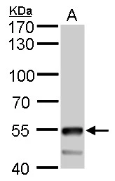
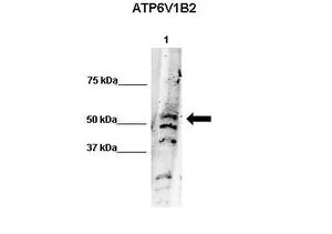
ATP6V1B2 antibody detects ATP6V1B2 protein by western blot analysis. A. 50 μg mouse brain lysate/extract 7.5 % SDS-PAGE ATP6V1B2 antibody (GTX110783) dilution: 1:5000
 dilution: 1:1000")
 dilution: 1:500.
Antigen Retrieval: Trilogy? (EDTA based, pH 8.0) buffer, 15min")
 dilution: 1:5000")
 in 2019.")
 in 2019.")
 in 2019.")
ATP6V1B2 antibody detects ATP6V1B2 protein by western blot analysis. A. 50 μg mouse brain lysate/extract 7.5 % SDS-PAGE ATP6V1B2 antibody (GTX110783) dilution: 1:5000
ATP6V1B2 antibody
GTX110783
ApplicationsWestern Blot, ImmunoHistoChemistry, ImmunoHistoChemistry Paraffin
Product group Antibodies
ReactivityHuman, Mouse, Rat
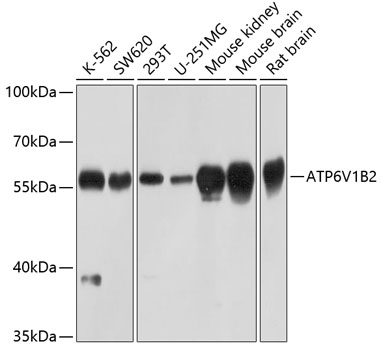
TargetATP6V1B2
Overview
- SupplierGeneTex
- Product NameATP6V1B2 antibody
- Delivery Days Customer9
- Application Supplier NoteWB: 1:500-1:10000. IHC-P: 1:100-1:1000. *Optimal dilutions/concentrations should be determined by the researcher.Not tested in other applications.
- ApplicationsWestern Blot, ImmunoHistoChemistry, ImmunoHistoChemistry Paraffin
- CertificationResearch Use Only
- ClonalityPolyclonal
- Concentration1 mg/ml
- ConjugateUnconjugated
- Gene ID526
- Target nameATP6V1B2
- Target descriptionATPase H+ transporting V1 subunit B2
- Target synonymsATP6B1B2, ATP6B2, DOOD, HO57, VATB, VPP3, Vma2, ZLS2, V-type proton ATPase subunit B, brain isoform, ATPase, H+ transporting, lysosomal 56/58kDa, V1 subunit B2, H+ transporting two-sector ATPase, V-ATPase B2 subunit, V-ATPase subunit B 2, endomembrane proton pump 58 kDa subunit, testicular secretory protein Li 65, vacuolar H+-ATPase 56,000 subunit, vacuolar proton pump subunit B 2
- HostRabbit
- IsotypeIgG
- Protein IDP21281
- Protein NameV-type proton ATPase subunit B, brain isoform
- Scientific DescriptionThis gene encodes a component of vacuolar ATPase (V-ATPase), a multisubunit enzyme that mediates acidification of eukaryotic intracellular organelles. V-ATPase dependent organelle acidification is necessary for such intracellular processes as protein sorting, zymogen activation, receptor-mediated endocytosis, and synaptic vesicle proton gradient generation. V-ATPase is composed of a cytosolic V1 domain and a transmembrane V0 domain. The V1 domain consists of three A, three B, and two G subunits, as well as a C, D, E, F, and H subunit. The V1 domain contains the ATP catalytic site. The protein encoded by this gene is one of two V1 domain B subunit isoforms and is the only B isoform highly expressed in osteoclasts. [provided by RefSeq, Jul 2008]
- ReactivityHuman, Mouse, Rat
- Storage Instruction-20°C or -80°C,2°C to 8°C
- UNSPSC41116161
Datasheet
Related products

Product group Antibodies
Anti-ATP6V1B2 Antibody Picoband(r)A04927-1-CARRIER-FREE
ApplicationsWestern Blot
ReactivityHuman, Mouse, Rat
TargetATP6V1B2
- SizePrice
Product group Antibodies
Anti-ATP6V1B2 AntibodyA17202
ApplicationsWestern Blot
ReactivityHuman, Mouse, Rat
- SizePrice
Product group Antibodies
Anti-ATP6V1B2 Antibody144-03754
ApplicationsWestern Blot
ReactivityHuman, Mouse, Rat
TargetATP6V1B2
- SizePrice
Product group Antibodies

ATP6V1B2 AntibodyLS-C668373
ApplicationsWestern Blot
ReactivityHuman
TargetATP6V1B2
- SizePrice
Product group Antibodies
ATP6V1B2 Polyclonal AntibodyBS-12549R
ApplicationsImmunoFluorescence, Western Blot, ELISA, ImmunoCytoChemistry, ImmunoHistoChemistry, ImmunoHistoChemistry Frozen, ImmunoHistoChemistry Paraffin
ReactivityBovine, Canine, Chicken, Equine, Human, Mouse, Porcine, Rabbit, Rat, Sheep
TargetATP6V1B2
- SizePrice
Product group Antibodies
ATP6V1B2 AntibodyCSB-PA002398ESR1HU
ApplicationsWestern Blot, ELISA
ReactivityHuman
TargetATP6V1B2
- SizePrice

Product group Antibodies
Anti-ATP6V1B2 AntibodyHPA008147
ApplicationsWestern Blot, ImmunoCytoChemistry, ImmunoHistoChemistry
ReactivityHuman, Mouse, Rat
TargetATP6V1B2
- SizePrice
Product group Antibodies
ATP6V1B2 antibody, InternalGTX45065
ApplicationsWestern Blot
ReactivityHuman
TargetATP6V1B2
- SizePrice
Product group Antibodies
ATP6V1B2 antibody, N-termGTX45066
ApplicationsWestern Blot
ReactivityHuman, Mouse
TargetATP6V1B2
- SizePrice

Product group Antibodies
ATP6V1B2 antibodyGTX32461
ApplicationsImmunoFluorescence, Western Blot, ImmunoCytoChemistry
ReactivityHuman, Mouse, Rat
TargetATP6V1B2
- SizePrice