

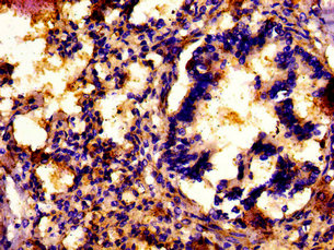
IHC image of CSB-PA863972LA01HU diluted at 1: 500 and staining in paraffin-embedded human lung tissue performed on a Leica BondTM system. After dewaxing and hydration, antigen retrieval was mediated by high pressure in a citrate buffer (pH 6.0) . Section was blocked with 10% normal goat serum 30min at RT. Then primary antibody (1% BSA) was incubated at 4°C overnight. The primary is detected by a biotinylated secondary antibody and visualized using an HRP conjugated SP system.
.")
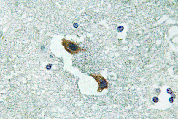
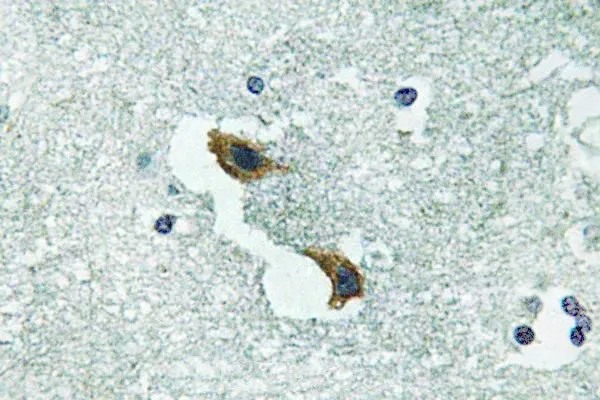
IHC image of CSB-PA863972LA01HU diluted at 1: 500 and staining in paraffin-embedded human lung tissue performed on a Leica BondTM system. After dewaxing and hydration, antigen retrieval was mediated by high pressure in a citrate buffer (pH 6.0) . Section was blocked with 10% normal goat serum 30min at RT. Then primary antibody (1% BSA) was incubated at 4°C overnight. The primary is detected by a biotinylated secondary antibody and visualized using an HRP conjugated SP system.
ATP7A Antibody
CSB-PA002414LA01HU
ApplicationsImmunoFluorescence, ELISA, ImmunoHistoChemistry
Product group Antibodies
ReactivityHuman
TargetATP7A
Overview
- SupplierCusabio
- Product NameATP7A Antibody
- Delivery Days Customer20
- ApplicationsImmunoFluorescence, ELISA, ImmunoHistoChemistry
- CertificationResearch Use Only
- ClonalityPolyclonal
- ConjugateUnconjugated
- Gene ID538
- Target nameATP7A
- Target descriptionATPase copper transporting alpha
- Target synonymsDSMAX, HMNX, MK, MNK, SMAX3, copper-transporting ATPase 1, ATPase, Cu++ transporting, alpha polypeptide, Cu++-transporting P-type ATPase, Menkes disease-associated protein, copper pump 1
- HostRabbit
- IsotypeIgG
- Protein IDQ04656
- Protein NameCopper-transporting ATPase 1
- Scientific DescriptionMay supply copper to copper-requiring proteins within the secretory pathway, when localized in the trans-Golgi network. Under conditions of elevated extracellular copper, it relocalized to the plasma membrane where it functions in the efflux of copper from cells.
- ReactivityHuman
- Storage Instruction-20°C or -80°C
- UNSPSC41116161
Related products
Product group Antibodies
Anti-ATP7A AntibodyA97049
ApplicationsELISA, ImmunoHistoChemistry
ReactivityHuman, Mouse, Rat
- SizePrice

Product group Antibodies
Anti-ATP7A Antibody Picoband(r)A01085-1-CARRIER-FREE
ApplicationsFlow Cytometry, Western Blot
ReactivityHuman
TargetATP7A
- SizePrice
Product group Antibodies

Anti-ATP7A [L60/4]AB02124-10.0-BT
ApplicationsImmunoPrecipitation, Western Blot, ImmunoHistoChemistry
ReactivityHuman, Mouse, Rat
TargetATP7A
- SizePrice

Product group Antibodies
Anti-ATP7A AntibodyHPA012887
ApplicationsImmunoHistoChemistry
ReactivityHuman
TargetATP7A
- SizePrice
Product group Antibodies

MNK / ATP7A AntibodyLS-C402996
ApplicationsWestern Blot, ELISA, ImmunoHistoChemistry
ReactivityHuman, Mouse, Rat
TargetATP7A
- SizePrice
Product group Antibodies
References
ATP7A Polyclonal AntibodyBS-1572R
ApplicationsImmunoFluorescence, Western Blot, ELISA, ImmunoCytoChemistry, ImmunoHistoChemistry, ImmunoHistoChemistry Frozen, ImmunoHistoChemistry Paraffin
ReactivityBovine, Canine, Equine, Human, Mouse, Rabbit, Rat
TargetATP7A
- SizePrice
Product group Antibodies
ATP7A antibodyGTX66723
ApplicationsImmunoHistoChemistry, ImmunoHistoChemistry Paraffin
ReactivityHuman
TargetATP7A
- SizePrice