

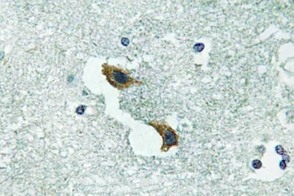
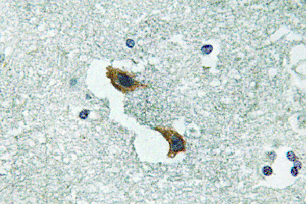
IHC-P analysis of human brain tissue using GTX66723 ATP7A antibody.
IHC-P analysis of human brain tissue using GTX66723 ATP7A antibody.
ATP7A antibody
GTX66723
ApplicationsImmunoHistoChemistry, ImmunoHistoChemistry Paraffin
Product group Antibodies
ReactivityHuman
TargetATP7A
Overview
- SupplierGeneTex
- Product NameATP7A antibody
- Delivery Days Customer9
- Application Supplier NoteIHC-P: 1:50-1:200. *Optimal dilutions/concentrations should be determined by the researcher.Not tested in other applications.
- ApplicationsImmunoHistoChemistry, ImmunoHistoChemistry Paraffin
- CertificationResearch Use Only
- ClonalityPolyclonal
- Concentration1 mg/ml
- ConjugateUnconjugated
- Gene ID538
- Target nameATP7A
- Target descriptionATPase copper transporting alpha
- Target synonymsDSMAX, HMNX, MK, MNK, SMAX3, copper-transporting ATPase 1, ATPase, Cu++ transporting, alpha polypeptide, Cu++-transporting P-type ATPase, Menkes disease-associated protein, copper pump 1
- HostRabbit
- IsotypeIgG
- Protein IDQ04656
- Protein NameCopper-transporting ATPase 1
- Scientific DescriptionThis gene encodes a transmembrane protein that functions in copper transport across membranes. This protein is localized to the trans Golgi network, where it is predicted to supply copper to copper-dependent enzymes in the secretory pathway. It relocalizes to the plasma membrane under conditions of elevated extracellular copper, and functions in the efflux of copper from cells. Mutations in this gene are associated with Menkes disease, X-linked distal spinal muscular atrophy, and occipital horn syndrome. Alternatively-spliced transcript variants have been observed. [provided by RefSeq, Aug 2013]
- ReactivityHuman
- Storage Instruction-20°C or -80°C,2°C to 8°C
- UNSPSC41116161
Datasheet
Related products
Product group Antibodies
Anti-ATP7A AntibodyA97049
ApplicationsELISA, ImmunoHistoChemistry
ReactivityHuman, Mouse, Rat
- SizePrice
Product group Antibodies

Anti-ATP7A [L60/4]AB02124-10.0-BT
ApplicationsImmunoPrecipitation, Western Blot, ImmunoHistoChemistry
ReactivityHuman, Mouse, Rat
TargetATP7A
- SizePrice

Product group Antibodies
Anti-ATP7A Antibody Picoband(r)A01085-1-CARRIER-FREE
ApplicationsFlow Cytometry, Western Blot
ReactivityHuman
TargetATP7A
- SizePrice
Product group Antibodies
ATP7A AntibodyCSB-PA002414LA01HU
ApplicationsImmunoFluorescence, ELISA, ImmunoHistoChemistry
ReactivityHuman
TargetATP7A
- SizePrice

Product group Antibodies
Anti-ATP7A AntibodyHPA012887
ApplicationsImmunoHistoChemistry
ReactivityHuman
TargetATP7A
- SizePrice
Product group Antibodies

MNK / ATP7A AntibodyLS-C402996
ApplicationsWestern Blot, ELISA, ImmunoHistoChemistry
ReactivityHuman, Mouse, Rat
TargetATP7A
- SizePrice
Product group Antibodies
References
ATP7A Polyclonal AntibodyBS-1572R
ApplicationsImmunoFluorescence, Western Blot, ELISA, ImmunoCytoChemistry, ImmunoHistoChemistry, ImmunoHistoChemistry Frozen, ImmunoHistoChemistry Paraffin
ReactivityBovine, Canine, Equine, Human, Mouse, Rabbit, Rat
TargetATP7A
- SizePrice