

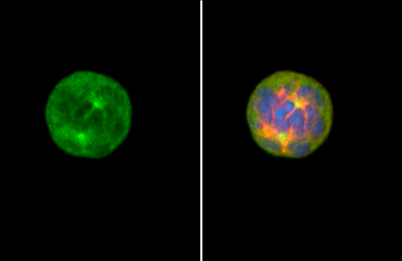
Aurora A antibody detects Aurora A protein at centrosome by immunofluorescent analysis. Sample: HeLa cells were fixed in 4% paraformaldehyde at RT for 15 min. Green: Aurora A stained by Aurora A antibody (GTX134467) diluted at 1:500. Red: alpha Tubulin, a cytoskeleton marker, stained by alpha Tubulin antibody [GT114] (GTX628802) diluted at 1:1000. Blue: Fluoroshield with DAPI (GTX30920).
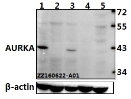
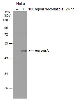
 and transfected (+) 293T whole cell extracts (30 μg) were separated by 10% SDS-PAGE, and the membrane was blotted with Aurora A antibody (GTX134467) diluted at 1:5000. The HRP-conjugated anti-rabbit IgG antibody (GTX213110-01) was used to detect the primary antibody.")
Aurora A antibody detects Aurora A protein at centrosome by immunofluorescent analysis. Sample: HeLa cells were fixed in 4% paraformaldehyde at RT for 15 min. Green: Aurora A stained by Aurora A antibody (GTX134467) diluted at 1:500. Red: alpha Tubulin, a cytoskeleton marker, stained by alpha Tubulin antibody [GT114] (GTX628802) diluted at 1:1000. Blue: Fluoroshield with DAPI (GTX30920).
Aurora A antibody
GTX134467
ApplicationsImmunoFluorescence, Western Blot, ImmunoCytoChemistry
Product group Antibodies
ReactivityHuman
TargetAURKA
Overview
- SupplierGeneTex
- Product NameAurora A antibody
- Delivery Days Customer9
- ApplicationsImmunoFluorescence, Western Blot, ImmunoCytoChemistry
- CertificationResearch Use Only
- ClonalityPolyclonal
- Concentration1.5 mg/ml
- ConjugateUnconjugated
- Gene ID6790
- Target nameAURKA
- Target descriptionaurora kinase A
- Target synonymsAIK, ARK1, AURA, BTAK, PPP1R47, STK15, STK6, STK7, aurora kinase A, aurora 2, aurora/IPL1-like kinase, aurora/IPL1-related kinase 1, breast tumor-amplified kinase, protein phosphatase 1, regulatory subunit 47, serine/threonine protein kinase 15, serine/threonine-protein kinase 6, serine/threonine-protein kinase aurora-A
- HostRabbit
- IsotypeIgG
- Protein IDO14965
- Protein NameAurora kinase A
- Scientific DescriptionThe protein encoded by this gene is a cell cycle-regulated kinase that appears to be involved in microtubule formation and/or stabilization at the spindle pole during chromosome segregation. The encoded protein is found at the centrosome in interphase cells and at the spindle poles in mitosis. This gene may play a role in tumor development and progression. A processed pseudogene of this gene has been found on chromosome 1, and an unprocessed pseudogene has been found on chromosome 10. Multiple transcript variants encoding the same protein have been found for this gene. [provided by RefSeq, Jul 2008]
- ReactivityHuman
- Storage Instruction-20°C or -80°C,2°C to 8°C
- UNSPSC41116161
Datasheet
Related products
Product group Antibodies
ApplicationsWestern Blot
ReactivityHuman, Mouse, Rat
- SizePrice
Product group Antibodies
Anti-AURKA Antibody144-02121
ApplicationsWestern Blot
ReactivityHuman, Mouse
TargetAURKA
- SizePrice
Product group Antibodies

AURKA / Aurora-A AntibodyLS-C831219
ApplicationsELISA, ImmunoHistoChemistry
ReactivityHuman
TargetAURKA
- SizePrice
Product group Antibodies
Aurora A Recombinant AntibodyBSM-52018R
ApplicationsImmunoFluorescence, ImmunoCytoChemistry, ImmunoHistoChemistry, ImmunoHistoChemistry Frozen, ImmunoHistoChemistry Paraffin
ReactivityHuman, Mouse, Rat
TargetAURKA
- SizePrice

Product group Antibodies
Anti-Aurora A/AURKA Antibody Picoband(r)A00246-3-CARRIER-FREE
ApplicationsFlow Cytometry, ImmunoFluorescence, Western Blot, ELISA, ImmunoCytoChemistry, ImmunoHistoChemistry
ReactivityHuman
TargetAURKA
- SizePrice
Product group Antibodies
AURKA AntibodyCSB-PA000924
ApplicationsWestern Blot, ELISA
ReactivityHuman, Mouse, Rat
TargetAURKA
- SizePrice
Product group Antibodies
References

ApplicationsImmunoPrecipitation, Western Blot, ELISA
ReactivityHuman
TargetAURKA
- SizePrice
Product group Antibodies
Aurka Polyclonal AntibodyCAC07174
ApplicationsImmunoFluorescence, Western Blot, ELISA, ImmunoHistoChemistry
ReactivityMouse
TargetAURKA
- SizePrice
Product group Antibodies
Aurora A antibodyGTX13408
ApplicationsImmunoPrecipitation, Western Blot
ReactivityHuman
TargetAURKA
- SizePrice
Product group Antibodies
Aurora A antibodyGTX134461
ApplicationsWestern Blot
ReactivityHuman
TargetAURKA
- SizePrice