

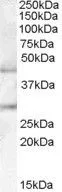
WB analysis of human pancreas lysate using GTX88963 B3GNT2 antibody, Internal. Dilution : 0.1μg/ml Loading : 35μg protein in RIPA buffer
WB analysis of human pancreas lysate using GTX88963 B3GNT2 antibody, Internal. Dilution : 0.1μg/ml Loading : 35μg protein in RIPA buffer
B3GNT2 antibody, Internal
GTX88963
ApplicationsWestern Blot
Product group Antibodies
ReactivityHuman
TargetB3GNT2
Overview
- SupplierGeneTex
- Product NameB3GNT2 antibody, Internal
- Delivery Days Customer7
- Application Supplier NoteWB: 0.1-0.3microg/ml. *Optimal dilutions/concentrations should be determined by the researcher.Not tested in other applications.
- ApplicationsWestern Blot
- CertificationResearch Use Only
- ClonalityPolyclonal
- Concentration0.50 mg/ml
- ConjugateUnconjugated
- Gene ID10678
- Target nameB3GNT2
- Target descriptionUDP-GlcNAc:betaGal beta-1,3-N-acetylglucosaminyltransferase 2
- Target synonyms3-Gn-T1, 3-Gn-T2, B3GN-T2, B3GNT, B3GNT-2, B3GNT1, BETA3GNT, BGNT2, BGnT-2, beta-1, beta3Gn-T1, beta3Gn-T2, N-acetyllactosaminide beta-1,3-N-acetylglucosaminyltransferase 2, UDP-GlcNAc:betaGal beta-1,3-N-acetylglucosaminyltransferase 1, beta-1,3-N-acetylglucosaminyltransferase bGnT-1, beta-1,3-N-acetylglucosaminyltransferase bGnT-2
- HostGoat
- IsotypeIgG
- Protein IDQ9NY97
- Protein NameN-acetyllactosaminide beta-1,3-N-acetylglucosaminyltransferase 2
- Scientific DescriptionThis gene encodes a member of the beta-1,3-N-acetylglucosaminyltransferase family. This enzyme is a type II transmembrane protein. It prefers the substrate of lacto-N-neotetraose, and is involved in the biosynthesis of poly-N-acetyllactosamine chains. Two transcript variants encoding the same protein have been found for this gene. [provided by RefSeq, Jan 2016]
- ReactivityHuman
- Storage Instruction-20°C or -80°C,2°C to 8°C
- UNSPSC41116161
Datasheet
Related products
Product group Antibodies
B3GNT2 AntibodyCSB-PA002500LA01HU
ApplicationsWestern Blot, ELISA, ImmunoHistoChemistry
ReactivityHuman
TargetB3GNT2
- SizePrice

Product group Antibodies
Anti-B3GNT2 Antibody Picoband(r)A09524-1-CARRIER-FREE
ApplicationsFlow Cytometry, ImmunoFluorescence, Western Blot, ELISA, ImmunoCytoChemistry
ReactivityHuman, Mouse, Rat
TargetB3GNT2
- SizePrice
Product group Antibodies

Goat anti-B3GNT2EB08038
ApplicationsWestern Blot, ELISA
ReactivityHuman, Mouse, Rat
TargetB3GNT2
- SizePrice

Product group Antibodies
Anti-B3GNT2-25ulHPA005997
ApplicationsWestern Blot, ImmunoCytoChemistry
ReactivityHuman
- SizePrice
Product group Antibodies

B3GNT2 AntibodyLS-C375293
ApplicationsELISA, ImmunoHistoChemistry, ImmunoHistoChemistry Paraffin
ReactivityHuman
TargetB3GNT2
- SizePrice
Product group Antibodies
B3Gnt2 Polyclonal AntibodyCAC07486
ApplicationsWestern Blot, ELISA, ImmunoHistoChemistry
TargetB3GNT2
- SizePrice