

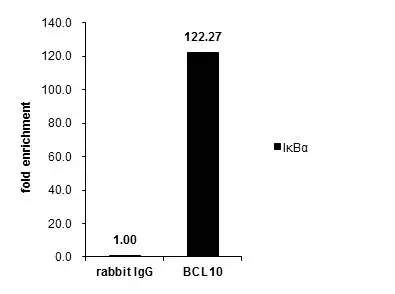
Cross-linked ChIP was performed with MCF-7 chromatin extract treated with TNF-a (10 ng/ml for 30 min) and 5 μg of either control rabbit IgG or anti-BCL10 antibody. The precipitated DNA was detected by PCR with primer set targeting to IκBα.
![BCL10 antibody [N1C3] detects BCL10 protein at cytoplasm on human colon carcinoma by immunohistochemical analysis. Sample: Paraffin-embedded human colon carcinoma. BCL10 antibody [N1C3] (GTX109159) diluted at 1:500.
Antigen Retrieval: Trilogy? (EDTA based, pH 8.0) buffer, 15min](https://www.genetex.com/upload/website/prouct_img/normal/GTX109159/GTX109159_39988_20141111_IHC_2_w_23060120_112.webp "BCL10 antibody [N1C3] detects BCL10 protein at cytoplasm on human colon carcinoma by immunohistochemical analysis. Sample: Paraffin-embedded human colon carcinoma. BCL10 antibody [N1C3] (GTX109159) diluted at 1:500.
Antigen Retrieval: Trilogy? (EDTA based, pH 8.0) buffer, 15min")
![BCL10 antibody [N1C3] detects BCL10 protein at cytoplasm on human breast carcinoma by immunohistochemical analysis. Sample: Paraffin-embedded human breast carcinoma. BCL10 antibody [N1C3] (GTX109159) diluted at 1:500.
Antigen Retrieval: Trilogy? (EDTA based, pH 8.0) buffer, 15min](https://www.genetex.com/upload/website/prouct_img/normal/GTX109159/GTX109159_39988_20141110_IHC_w_23060120_281.webp "BCL10 antibody [N1C3] detects BCL10 protein at cytoplasm on human breast carcinoma by immunohistochemical analysis. Sample: Paraffin-embedded human breast carcinoma. BCL10 antibody [N1C3] (GTX109159) diluted at 1:500.
Antigen Retrieval: Trilogy? (EDTA based, pH 8.0) buffer, 15min")
![BCL10 antibody [N1C3] immunoprecipitates BCL10 protein in IP experiments. IP samples: Raji whole cell extract A. Control with 4 μg of preimmune Rabbit IgG B. Immunoprecipitation of BCL10 protein by 4 μg BCL10 antibody [N1C3] (GTX109159) 12 % SDS-PAGE The immunoprecipitated BCL10 protein was detected by BCL10 antibody [N1C3] (GTX109159) diluted at 1:500. [EasyBlot anti-rabbit IgG (GTX221666-01) was used as a secondary reagent]](https://www.genetex.com/upload/website/prouct_img/normal/GTX109159/GTX109159_39988_IP_w_23060120_533.webp "BCL10 antibody [N1C3] immunoprecipitates BCL10 protein in IP experiments. IP samples: Raji whole cell extract A. Control with 4 μg of preimmune Rabbit IgG B. Immunoprecipitation of BCL10 protein by 4 μg BCL10 antibody [N1C3] (GTX109159) 12 % SDS-PAGE The immunoprecipitated BCL10 protein was detected by BCL10 antibody [N1C3] (GTX109159) diluted at 1:500. [EasyBlot anti-rabbit IgG (GTX221666-01) was used as a secondary reagent]")
 A: NIH-3T3 12% SDS PAGE BCL10 antibody GTX109159 diluted at 1:1000")
![Various whole cell extracts (30 μg) were separated by 12% SDS-PAGE, and the membrane was blotted with BCL10 antibody [N1C3] (GTX109159) diluted at 1:1000. The HRP-conjugated anti-rabbit IgG antibody (GTX213110-01) was used to detect the primary antibody. Corresponding RNA expression data for the same cell lines are based on Human Protein Atlas program.](https://www.genetex.com/upload/website/prouct_img/normal/GTX109159/GTX109159_39988_20231208_WB_TPM_watermark_23121122_874.webp "Various whole cell extracts (30 μg) were separated by 12% SDS-PAGE, and the membrane was blotted with BCL10 antibody [N1C3] (GTX109159) diluted at 1:1000. The HRP-conjugated anti-rabbit IgG antibody (GTX213110-01) was used to detect the primary antibody. Corresponding RNA expression data for the same cell lines are based on Human Protein Atlas program.")
![Various whole cell extracts (30 μg) were separated by 12% SDS-PAGE, and the membrane was blotted with BCL10 antibody [N1C3] (GTX109159) diluted at 1:1000. The HRP-conjugated anti-rabbit IgG antibody (GTX213110-01) was used to detect the primary antibody, and the signal was developed with Trident ECL plus-Enhanced. Corresponding RNA expression data for the same cell lines are based on Human Protein Atlas program.](https://www.genetex.com/upload/website/prouct_img/normal/GTX109159/GTX109159_39988_20231208_WB_TPM_2_watermark_24032617_793.webp "Various whole cell extracts (30 μg) were separated by 12% SDS-PAGE, and the membrane was blotted with BCL10 antibody [N1C3] (GTX109159) diluted at 1:1000. The HRP-conjugated anti-rabbit IgG antibody (GTX213110-01) was used to detect the primary antibody, and the signal was developed with Trident ECL plus-Enhanced. Corresponding RNA expression data for the same cell lines are based on Human Protein Atlas program.")
Cross-linked ChIP was performed with MCF-7 chromatin extract treated with TNF-a (10 ng/ml for 30 min) and 5 μg of either control rabbit IgG or anti-BCL10 antibody. The precipitated DNA was detected by PCR with primer set targeting to IκBα.
BCL10 antibody [N1C3]
GTX109159
ApplicationsImmunoPrecipitation, Western Blot, ChIP Chromatin ImmunoPrecipitation, ImmunoHistoChemistry, ImmunoHistoChemistry Paraffin
Product group Antibodies
ReactivityHuman, Mouse
TargetBCL10
Overview
- SupplierGeneTex
- Product NameBCL10 antibody [N1C3]
- Delivery Days Customer9
- Application Supplier NoteWB: 1:500-1:3000. IHC-P: 1:100-1:1000. IP: 1:100-1:500. *Optimal dilutions/concentrations should be determined by the researcher.Not tested in other applications.
- ApplicationsImmunoPrecipitation, Western Blot, ChIP Chromatin ImmunoPrecipitation, ImmunoHistoChemistry, ImmunoHistoChemistry Paraffin
- CertificationResearch Use Only
- ClonalityPolyclonal
- Concentration0.45 mg/ml
- ConjugateUnconjugated
- Gene ID8915
- Target nameBCL10
- Target descriptionBCL10 immune signaling adaptor
- Target synonymsCARMEN, CIPER, CLAP, IMD37, c-E10, mE10, B-cell lymphoma/leukemia 10, B cell CLL/lymphoma 10, CARD containing molecule enhancing NF-kB, CARD-containing apoptotic signaling protein, CARD-containing molecule enhancing NF-kappa-B, CARD-containing proapoptotic protein, CED-3/ICH-1 prodomain homologous E10-like regulator, cCARMEN, caspase-recruiting domain-containing protein, cellular homolog of vCARMEN, cellular-E10, hCLAP, mammalian CARD-containing adapter molecule E10
- HostRabbit
- IsotypeIgG
- Protein IDO95999
- Protein NameB-cell lymphoma/leukemia 10
- Scientific DescriptionThis gene was identified by its translocation in a case of mucosa-associated lymphoid tissue (MALT) lymphoma. The protein encoded by this gene contains a caspase recruitment domain (CARD), and has been shown to induce apoptosis and to activate NF-kappaB. This protein is reported to interact with other CARD domain containing proteins including CARD9, 10, 11 and 14, which are thought to function as upstream regulators in NF-kappaB signaling. This protein is found to form a complex with MALT1, a protein encoded by another gene known to be translocated in MALT lymphoma. MALT1 and this protein are thought to synergize in the activation of NF-kappaB, and the deregulation of either of them may contribute to the same pathogenetic process that leads to the malignancy. [provided by RefSeq]
- ReactivityHuman, Mouse
- Storage Instruction-20°C or -80°C,2°C to 8°C
- UNSPSC41116161
Datasheet
Related products
Product group Antibodies
Anti-BCL10 AntibodyA95930
ApplicationsWestern Blot, ELISA, ImmunoHistoChemistry
ReactivityHuman, Mouse, Rat
- SizePrice
Product group Antibodies
Anti-BCL10 Antibody144-01106
ApplicationsWestern Blot
ReactivityHuman
TargetBCL10
- SizePrice
Product group Antibodies
ApplicationsFlow Cytometry, Western Blot, ImmunoCytoChemistry
ReactivityHuman
TargetBCL10
- SizePrice

Product group Antibodies
BCL10 AntibodyCSB-PA002608ESR2HU
ApplicationsWestern Blot, ELISA, ImmunoHistoChemistry
ReactivityHuman, Mouse
TargetBCL10
- SizePrice
Product group Antibodies
Bcl10 Polyclonal AntibodyCAC07487
ApplicationsImmunoFluorescence, ELISA, ImmunoHistoChemistry
TargetBCL10
- SizePrice
Product group Antibodies

BCL10 / BCL-10 AntibodyLS-C401444
ApplicationsWestern Blot, ELISA, ImmunoHistoChemistry
ReactivityHuman, Mouse, Rat
TargetBCL10
- SizePrice
![ICC/IF analysis of PFA-fixed K562 cells using GTX02597 BCL10 antibody [BL10/2988R].](https://www.genetex.com/upload/website/prouct_img/normal/GTX02597/GTX02597_20210319_ICCIF_w_23053122_353.webp)
Product group Antibodies
BCL10 antibody [BL10/2988R]GTX02597
ApplicationsFlow Cytometry, ImmunoFluorescence, ImmunoCytoChemistry, ImmunoHistoChemistry, ImmunoHistoChemistry Paraffin
ReactivityHuman
TargetBCL10
- SizePrice
![IHC-P analysis of human tonsil tissue using GTX04716 BCL10 antibody [151]. Antigen retrieval : EDTA Buffer pH 8.0](https://www.genetex.com/upload/website/prouct_img/normal/GTX04716/GTX04716_20240325_IHC-P_24032422_706.webp)
Product group Antibodies
BCL10 antibody [151]GTX04716
ApplicationsImmunoHistoChemistry, ImmunoHistoChemistry Paraffin
ReactivityHuman
TargetBCL10
- SizePrice
![FACS analysis of Jurkat cells using GTX84835 BCL10 antibody [4A8]. Red : Primary antibody Blue : Negative control antibody](https://www.genetex.com/upload/website/prouct_img/normal/GTX84835/GTX84835_576_FACS_w_23061420_524.webp)
Product group Antibodies
BCL10 antibody [4A8]GTX84835
ApplicationsFlow Cytometry, ImmunoFluorescence, Western Blot, ImmunoCytoChemistry, ImmunoHistoChemistry, ImmunoHistoChemistry Paraffin
ReactivityHuman
TargetBCL10
- SizePrice
Product group Antibodies
BCL10 antibodyGTX112744
ApplicationsImmunoFluorescence, ImmunoPrecipitation, Western Blot, ImmunoCytoChemistry, ImmunoHistoChemistry, ImmunoHistoChemistry Paraffin
ReactivityHuman, Mouse
TargetBCL10
- SizePrice