

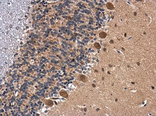
BDNF antibody detects BDNF protein at cytoplasm in rat brain by immunohistochemical analysis. Sample: Paraffin-embedded rat brain. BDNF antibody (GTX132621) diluted at 1:500.
Antigen Retrieval: Citrate buffer, pH 6.0, 15 min
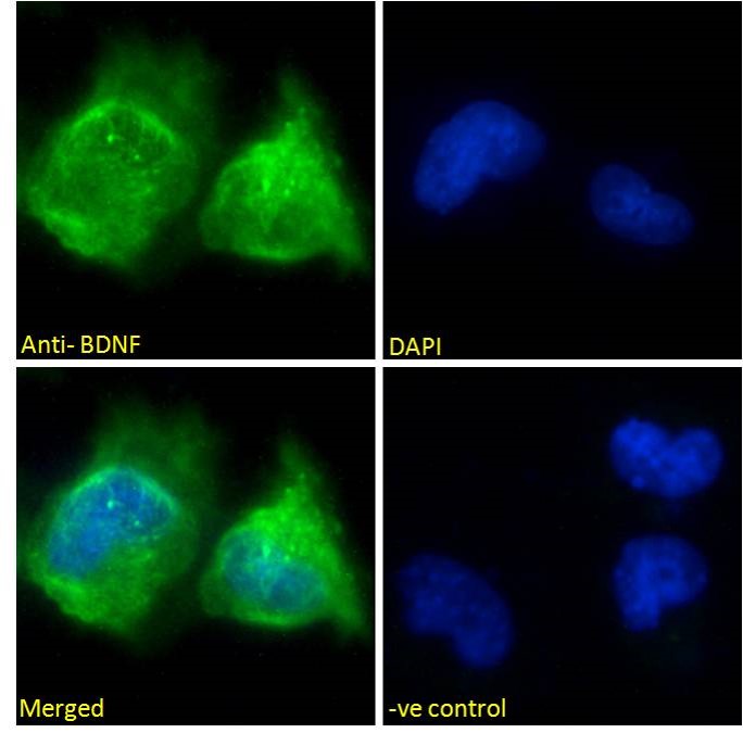
![BDNF antibody detects BDNF protein by immunofluorescent analysis. Sample: DIV10 rat E18 primary cortical neuron cells were fixed in 4% paraformaldehyde at RT for 15 min. Green: BDNF stained by BDNF antibody (GTX132621) diluted at 1:500. Red: Tau, stained by Tau antibody [GT287] (GTX634809) diluted at 1:500. Blue: Fluoroshield with DAPI (GTX30920).](https://www.genetex.com/upload/website/prouct_img/normal/GTX132621/GTX132621_43552_20191125_ICC_IF_R_w_23060523_650.webp "BDNF antibody detects BDNF protein by immunofluorescent analysis. Sample: DIV10 rat E18 primary cortical neuron cells were fixed in 4% paraformaldehyde at RT for 15 min. Green: BDNF stained by BDNF antibody (GTX132621) diluted at 1:500. Red: Tau, stained by Tau antibody [GT287] (GTX634809) diluted at 1:500. Blue: Fluoroshield with DAPI (GTX30920).")
 diluted at 1:1000. Antigen Retrieval: Citrate buffer, pH 6.0, 15 min")
 diluted at 1:500. Antigen Retrieval: Citrate buffer, pH 6.0, 15 min")
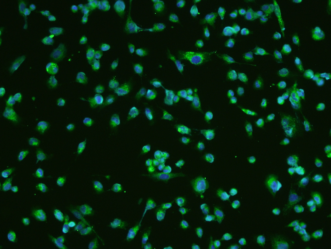
![BDNF antibody detects BDNF protein expression by immunohistochemical analysis. Sample:Paraffin-Embedded adult mouse retina. Green: BDNF protein stained by BDNF antibody (GTX132621) diluted at 1:250. Red: beta Tubulin 3/ TUJ1, stained by beta Tubulin 3/ TUJ1 antibody [GT11710] (GTX631836) diluted at 1:500. Blue: Fluoroshield with DAPI (GTX30920).](https://www.genetex.com/upload/website/prouct_img/normal/GTX132621/GTX132621_42529_20170522_IHC-P_M_w_23060523_250.webp "BDNF antibody detects BDNF protein expression by immunohistochemical analysis. Sample:Paraffin-Embedded adult mouse retina. Green: BDNF protein stained by BDNF antibody (GTX132621) diluted at 1:250. Red: beta Tubulin 3/ TUJ1, stained by beta Tubulin 3/ TUJ1 antibody [GT11710] (GTX631836) diluted at 1:500. Blue: Fluoroshield with DAPI (GTX30920).")
 were separated by 15% SDS-PAGE, and the membrane was blotted with BDNF antibody (GTX132621) diluted at 1:500. The HRP-conjugated anti-rabbit IgG antibody (GTX213110-01) was used to detect the primary antibody.")
 were separated by 15% SDS-PAGE, and the membrane was blotted with BDNF antibody (GTX132621) diluted at 1:125. The HRP-conjugated anti-rabbit IgG antibody (GTX213110-01) was used to detect the primary antibody.")
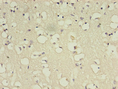
BDNF antibody detects BDNF protein at cytoplasm in rat brain by immunohistochemical analysis. Sample: Paraffin-embedded rat brain. BDNF antibody (GTX132621) diluted at 1:500.
Antigen Retrieval: Citrate buffer, pH 6.0, 15 min
BDNF antibody
GTX132621
ApplicationsImmunoFluorescence, Western Blot, ImmunoCytoChemistry, ImmunoHistoChemistry, ImmunoHistoChemistry Frozen, ImmunoHistoChemistry Paraffin
Product group Antibodies
ReactivityHuman, Mouse, Rat
TargetBDNF
Overview
- SupplierGeneTex
- Product NameBDNF antibody
- Delivery Days Customer9
- Application Supplier NoteWB: 1:500-1:3000. ICC/IF: 1:100-1:1000. IHC-P: 1:100-1:1000. *Optimal dilutions/concentrations should be determined by the researcher.Not tested in other applications.
- ApplicationsImmunoFluorescence, Western Blot, ImmunoCytoChemistry, ImmunoHistoChemistry, ImmunoHistoChemistry Frozen, ImmunoHistoChemistry Paraffin
- CertificationResearch Use Only
- ClonalityPolyclonal
- Concentration1.96 mg/ml
- ConjugateUnconjugated
- Gene ID627
- Target nameBDNF
- Target descriptionbrain derived neurotrophic factor
- Target synonymsANON2, BULN2, neurotrophic factor BDNF precursor form, abrineurin, neurotrophin, proBDNF
- HostRabbit
- IsotypeIgG
- Protein IDP23560
- Protein NameNeurotrophic factor BDNF precursor form
- Scientific DescriptionThe protein encoded by this gene is a member of the nerve growth factor family. It is induced by cortical neurons, and is necessary for survival of striatal neurons in the brain. Expression of this gene is reduced in both Alzheimers and Huntington disease patients. This gene may play a role in the regulation of stress response and in the biology of mood disorders. Multiple transcript variants encoding distinct isoforms have been described for this gene. [provided by RefSeq]
- ReactivityHuman, Mouse, Rat
- Storage Instruction-20°C or -80°C,2°C to 8°C
- UNSPSC41116161
Datasheet
Related products
Product group Antibodies
Anti-BDNF AntibodyA83462
ApplicationsImmunoFluorescence, ELISA
ReactivityHuman
- SizePrice
Product group Antibodies

Peroxiredoxin 3 (PRDX3) AntibodyABX329679
ApplicationsWestern Blot, ELISA, ImmunoHistoChemistry
- SizePrice

Product group Antibodies
References
ApplicationsImmunoFluorescence, Western Blot, ImmunoCytoChemistry, ImmunoHistoChemistry
ReactivityHuman, Mouse, Rat
TargetBDNF
- SizePrice
Product group Antibodies

BDNF AntibodyLS-C830275
ApplicationsWestern Blot, ELISA, ImmunoHistoChemistry
ReactivityHuman, Mouse, Rat
TargetBDNF
- SizePrice
Product group Antibodies
References
BDNF Polyclonal AntibodyBS-4989R
ApplicationsFlow Cytometry, ImmunoFluorescence, Western Blot, ELISA, ImmunoCytoChemistry, ImmunoHistoChemistry, ImmunoHistoChemistry Frozen, ImmunoHistoChemistry Paraffin
ReactivityHuman, Mouse, Rat
TargetBDNF
- SizePrice
Product group Antibodies
BDNF AntibodyCSB-PA002655LA01HU
ApplicationsELISA, ImmunoHistoChemistry
ReactivityHuman
TargetBDNF
- SizePrice
Product group Antibodies

Goat anti-BDNFEB08117
ApplicationsImmunoFluorescence, ELISA
ReactivityCanine, Human, Mouse, Rat
TargetBDNF
- SizePrice
Product group Antibodies
Bdnf Polyclonal AntibodyCAC07061
ApplicationsWestern Blot, ELISA, ImmunoHistoChemistry
ReactivityMouse
TargetBDNF
- SizePrice
Product group Antibodies
BDNF antibodyGTX17884
ApplicationsELISA
ReactivityHuman, Mouse
TargetBDNF
- SizePrice