


IHC-P analysis of mouse eye tissue using GTX14928 Bestrophin antibody. Dilution : 1:50
IHC-P analysis of mouse eye tissue using GTX14928 Bestrophin antibody. Dilution : 1:50
Bestrophin antibody
GTX14928
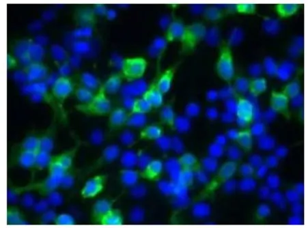
ApplicationsImmunoFluorescence, ImmunoPrecipitation, Western Blot, ELISA, ImmunoCytoChemistry, ImmunoHistoChemistry, ImmunoHistoChemistry Paraffin
Product group Antibodies
ReactivityHuman, Monkey, Mouse, Rat
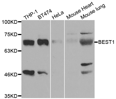
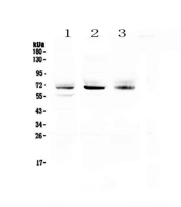
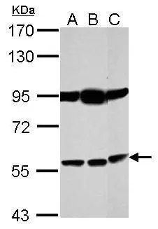
TargetBEST1
Overview
- SupplierGeneTex
- Product NameBestrophin antibody
- Delivery Days Customer9
- Application Supplier NoteWB: 1:500. ICC/IF: 1:100. IP: 1:200. ELISA: 1:10000. *Optimal dilutions/concentrations should be determined by the researcher.Not tested in other applications.
- ApplicationsImmunoFluorescence, ImmunoPrecipitation, Western Blot, ELISA, ImmunoCytoChemistry, ImmunoHistoChemistry, ImmunoHistoChemistry Paraffin
- CertificationResearch Use Only
- ClonalityPolyclonal
- Concentration0.65 mg/ml
- ConjugateUnconjugated
- Gene ID7439
- Target nameBEST1
- Target descriptionbestrophin 1
- Target synonymsARB, BEST, BMD, Best1V1Delta2, RP50, TU15B, VMD2, bestrophin-1, Best disease, vitelliform macular dystrophy protein 2
- HostRabbit
- IsotypeIgG
- Protein IDO76090
- Protein NameBestrophin-1
- Scientific DescriptionThis gene encodes a member of the bestrophin gene family. This small gene family is characterized by proteins with a highly conserved N-terminus with four to six transmembrane domains. Bestrophins may form chloride ion channels or may regulate voltage-gated L-type calcium-ion channels. Bestrophins are generally believed to form calcium-activated chloride-ion channels in epithelial cells but they have also been shown to be highly permeable to bicarbonate ion transport in retinal tissue. Mutations in this gene are responsible for juvenile-onset vitelliform macular dystrophy (VMD2), also known as Best macular dystrophy, in addition to adult-onset vitelliform macular dystrophy (AVMD) and other retinopathies. Alternative splicing results in multiple variants encoding distinct isoforms.[provided by RefSeq, Nov 2008]
- ReactivityHuman, Monkey, Mouse, Rat
- Storage Instruction-20°C or -80°C,2°C to 8°C
- UNSPSC41116161
Datasheet
Related products
Product group Antibodies
Anti-BEST1 AntibodyA31015
ApplicationsWestern Blot, ImmunoHistoChemistry
ReactivityHuman, Mouse
- SizePrice
Product group Antibodies
Anti-BEST1 Antibody144-03022
ApplicationsWestern Blot, ImmunoHistoChemistry
ReactivityHuman, Mouse, Rat
TargetBEST1
- SizePrice
Product group Antibodies

BEST1 / BEST / Bestrophin AntibodyLS-C749082
ApplicationsImmunoFluorescence, Western Blot
ReactivityHuman, Mouse
TargetBEST1
- SizePrice
Product group Antibodies
Anti-Bestrophin/BEST1 Antibody Picoband(r)A01434-1-CARRIER-FREE
ApplicationsFlow Cytometry, Western Blot, ImmunoCytoChemistry, ImmunoHistoChemistry
ReactivityHuman
TargetBEST1
- SizePrice
Product group Antibodies
BEST1 AntibodyCSB-PA080283
ApplicationsWestern Blot
ReactivityHuman, Mouse, Rat
TargetBEST1
- SizePrice
Product group Antibodies
ApplicationsImmunoPrecipitation, Western Blot, ImmunoCytoChemistry, ImmunoHistoChemistry
ReactivityPorcine
TargetBEST1
- SizePrice
Product group Antibodies
References
Bestrophin antibodyGTX14927
ApplicationsImmunoFluorescence, ImmunoPrecipitation, Western Blot, ELISA, ImmunoCytoChemistry, ImmunoHistoChemistry, ImmunoHistoChemistry Frozen, ImmunoHistoChemistry Paraffin
ReactivityHuman, Mouse, Rat
TargetBEST1
- SizePrice
![WB analysis of human RPE cell lysate using GTX30219 Bestrophin antibody[E6-6].](https://www.genetex.com/upload/website/prouct_img/normal/GTX30219/GTX30219_1367_WB_w_23060722_540.webp)
Product group Antibodies
Bestrophin antibody [E6-6]GTX30219
ApplicationsImmunoFluorescence, ImmunoPrecipitation, Western Blot, ImmunoCytoChemistry, ImmunoHistoChemistry, ImmunoHistoChemistry Frozen, ImmunoHistoChemistry Paraffin
ReactivityCanine, Human, Porcine, Primate
TargetBEST1
- SizePrice

Product group Antibodies
Anti-BEST1 AntibodyHPA057464
ApplicationsImmunoHistoChemistry
ReactivityHuman
TargetBEST1
- SizePrice
Product group Antibodies
Bestrophin 1 antibodyGTX105387
ApplicationsWestern Blot
ReactivityHuman
TargetBEST1
- SizePrice