

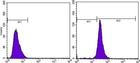
FACS analysis of MCF-7 cells using GTX83163 beta Actin antibody [8H10D10]. Right : beta Actin Left : negative control
![ICC/IF analysis of SKBR-3 (left) and A549 (right) cells using GTX83163 beta Actin antibody [8H10D10]. Red : beta Actin Blue: DRAQ5 fluorescent DNA dye](https://www.genetex.com/upload/website/prouct_img/normal/GTX83163/GTX83163_20170912_ICCIF_w_23061322_802.webp "ICC/IF analysis of SKBR-3 (left) and A549 (right) cells using GTX83163 beta Actin antibody [8H10D10]. Red : beta Actin Blue: DRAQ5 fluorescent DNA dye")
![WB analysis of NIH3T3 (1), Jurkat (2), HeLa (3), CHO (4), PC-12 (5), HEK293 (6), COS (7), A549 (8) and MCF-7 (9) cell lysate using GTX83163 beta Actin antibody [8H10D10].](https://www.genetex.com/upload/website/prouct_img/normal/GTX83163/GTX83163_20170912_WB_w_23061322_448.webp "WB analysis of NIH3T3 (1), Jurkat (2), HeLa (3), CHO (4), PC-12 (5), HEK293 (6), COS (7), A549 (8) and MCF-7 (9) cell lysate using GTX83163 beta Actin antibody [8H10D10].")
FACS analysis of MCF-7 cells using GTX83163 beta Actin antibody [8H10D10]. Right : beta Actin Left : negative control
beta Actin antibody [8H10D10]
GTX83163
ApplicationsFlow Cytometry, ImmunoFluorescence, Western Blot, ELISA, ImmunoCytoChemistry
Product group Antibodies
ReactivityHamster, Human, Monkey, Mouse, Rat
TargetACTB
Overview
- SupplierGeneTex
- Product Namebeta Actin antibody [8H10D10]
- Delivery Days Customer9
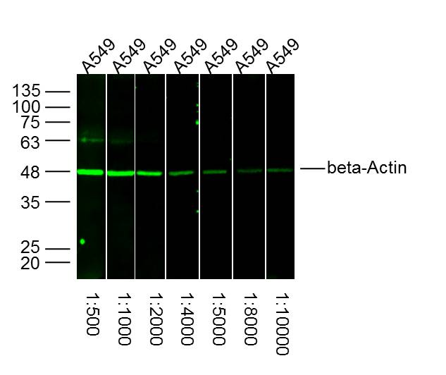
- Application Supplier NoteWB: 1/500 - 1/2000. ICC/IF: 1/200 - 1/1000. FACS: 1/200 - 1/400. ELISA: 1/10000. *Optimal dilutions/concentrations should be determined by the researcher.Not tested in other applications.
- ApplicationsFlow Cytometry, ImmunoFluorescence, Western Blot, ELISA, ImmunoCytoChemistry
- CertificationResearch Use Only
- ClonalityMonoclonal
- Clone ID8H10D10
- ConjugateUnconjugated
- Gene ID60
- Target nameACTB
- Target descriptionactin beta
- Target synonymsBKRNS, BNS, BRWS1, CSMH, DDS1, PS1TP5BP1, THC8, actin, cytoplasmic 1, I(2)-actin, PS1TP5-binding protein 1, beta cytoskeletal actin
- HostMouse
- IsotypeIgG2b
- Protein IDP60709
- Protein NameActin, cytoplasmic 1
- Scientific DescriptionThis gene encodes one of six different actin proteins. Actins are highly conserved proteins that are involved in cell motility, structure, and integrity. This actin is a major constituent of the contractile apparatus and one of the two nonmuscle cytoskeletal actins. [provided by RefSeq, Jul 2008]
- ReactivityHamster, Human, Monkey, Mouse, Rat
- Storage Instruction-20°C or -80°C,2°C to 8°C
- UNSPSC12352203
References
- Duan L, Pang HL, Chen WJ, et al. The role of GDF15 in bone metastasis of lung adenocarcinoma cells. Oncol Rep. 2019,41(4):2379-2388. doi: 10.3892/or.2019.7024Read this paper
Datasheet
Related products
Product group Antibodies
Actb Polyclonal AntibodyCAC07011
ApplicationsImmunoFluorescence, Western Blot, ELISA, ImmunoHistoChemistry
TargetACTB
- SizePrice
Product group Antibodies
References
beta-Actin Polyclonal AntibodyBS-0061R
ApplicationsFlow Cytometry, ImmunoFluorescence, Western Blot, ELISA, ImmunoCytoChemistry, ImmunoHistoChemistry, ImmunoHistoChemistry Frozen, ImmunoHistoChemistry Paraffin
ReactivityCanine, Chicken, Feline, Fish, Guinea Pig, Hamster, Human, Insect, Mouse, Porcine, Rabbit, Rat, Sheep
TargetACTB
- SizePrice
Product group Antibodies
Anti-beta Actin Antibody102-26634
ApplicationsWestern Blot
TargetACTB
- SizePrice
Product group Antibodies
Anti-beta Actin AntibodyA121645
ApplicationsImmunoFluorescence, Western Blot
ReactivityCanine, Human, Monkey, Mouse, Rat
- SizePrice

Product group Antibodies
Anti-ACTB AntibodyAMAB91241
ApplicationsWestern Blot, ImmunoCytoChemistry, ImmunoHistoChemistry
ReactivityHuman, Mouse, Rat
TargetACTB
- SizePrice
Product group Antibodies

Actin-pan AntibodyABX013019
ApplicationsWestern Blot, ELISA, ImmunoHistoChemistry
- SizePrice
Product group Antibodies

Anti-Beta-actin [Fab 19, actin]Ab02221-1.1
ApplicationsImmunoFluorescence, ELISA
ReactivityHuman
TargetACTB
- SizePrice
Product group Antibodies
References
F-Actin antibody [NH3]GTX76100
ApplicationsFlow Cytometry, ImmunoFluorescence, Western Blot, ELISA, ImmunoCytoChemistry, ImmunoHistoChemistry, ImmunoHistoChemistry Frozen
ReactivityHuman, Mouse, Rabbit, Rat
TargetACTB
- SizePrice
Product group Antibodies
References
Actin antibody [BA3R]GTX82559
ApplicationsImmunoFluorescence, Western Blot, ELISA, ImmunoCytoChemistry, ImmunoHistoChemistry
ReactivityChicken, Human, Mouse, Rabbit, Rat
TargetACTB
- SizePrice