

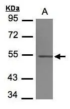
Sample(30 μg of whole cell lysate) A:293T 7.5% SDS PAGE GTX102095 diluted at 1:1000 The HRP-conjugated anti-rabbit IgG antibody (GTX213110-01) was used to detect the primary antibody.
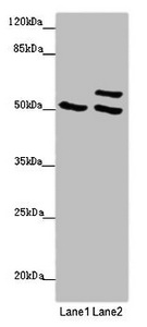
 A: NIH-3T3 10% SDS PAGE GTX102095 diluted at 1:1000 The HRP-conjugated anti-rabbit IgG antibody (GTX213110-01) was used to detect the primary antibody.")
 of paraformaldehyde-fixed U2OS, using beta Tubulin 4(GTX102095) antibody (Green) at 1:500 dilution. Alpha-tubulin filaments were labeled with GTX11304 (Red) at 1:2000.")
![beta Tubulin 4 antibody detects beta Tubulin 4 protein by immunofluorescent analysis. Sample: DIV9 rat E18 primary cortical neuron cells were fixed in 4% paraformaldehyde at RT for 15 min. Green: beta Tubulin 4 stained by beta Tubulin 4 antibody (GTX102095) diluted at 1:500. Red: beta Tubulin 3/ Tuj1, stained by beta Tubulin 3/ Tuj1 antibody [GT11710] (GTX631836) diluted at 1:500. Blue: Fluoroshield with DAPI (GTX30920).](https://www.genetex.com/upload/website/prouct_img/normal/GTX102095/GTX102095_39827_20181115_ICC_IF_R_w_23060100_942.webp "beta Tubulin 4 antibody detects beta Tubulin 4 protein by immunofluorescent analysis. Sample: DIV9 rat E18 primary cortical neuron cells were fixed in 4% paraformaldehyde at RT for 15 min. Green: beta Tubulin 4 stained by beta Tubulin 4 antibody (GTX102095) diluted at 1:500. Red: beta Tubulin 3/ Tuj1, stained by beta Tubulin 3/ Tuj1 antibody [GT11710] (GTX631836) diluted at 1:500. Blue: Fluoroshield with DAPI (GTX30920).")
Sample(30 μg of whole cell lysate) A:293T 7.5% SDS PAGE GTX102095 diluted at 1:1000 The HRP-conjugated anti-rabbit IgG antibody (GTX213110-01) was used to detect the primary antibody.
beta Tubulin 4 antibody
GTX102095
ApplicationsImmunoFluorescence, Western Blot, ImmunoCytoChemistry
Product group Antibodies
ReactivityHuman, Mouse, Rat
TargetTUBB4A
Overview
- SupplierGeneTex
- Product Namebeta Tubulin 4 antibody
- Delivery Days Customer9
- Application Supplier NoteWB: 1:500-1:3000. ICC/IF: 1:100-1:1000. *Optimal dilutions/concentrations should be determined by the researcher.Not tested in other applications.
- ApplicationsImmunoFluorescence, Western Blot, ImmunoCytoChemistry
- CertificationResearch Use Only
- ClonalityPolyclonal
- Concentration0.41 mg/ml
- ConjugateUnconjugated
- Gene ID10382
- Target nameTUBB4A
- Target descriptiontubulin beta 4A class IVa
- Target synonymsDYT4, TUBB4, beta-5, tubulin beta-4A chain, dystonia 4, torsion (autosomal dominant), tubulin beta-4 chain, tubulin, beta 4, tubulin, beta, 5
- HostRabbit
- IsotypeIgG
- Protein IDP04350
- Protein NameTubulin beta-4A chain
- Scientific DescriptionTubulin is the major constituent of microtubules. It binds two moles of GTP, one at an exchangeable site on the beta chain and one at a non-exchangeable site on the alpha-chain.
- ReactivityHuman, Mouse, Rat
- Storage Instruction-20°C or -80°C,2°C to 8°C
- UNSPSC41116161
Datasheet
Related products
Product group Antibodies
ApplicationsWestern Blot, ELISA
ReactivityHuman, Mouse
TargetTUBB4A
- SizePrice
Product group Antibodies
Anti-beta Tubulin 4 Antibody107-10316
ApplicationsImmunoFluorescence, Western Blot, ImmunoCytoChemistry
ReactivityHuman, Mouse
TargetTUBB4A
- SizePrice
Product group Antibodies

TUBB4 / Tubulin Beta 4 AntibodyLS-C679924
ApplicationsImmunoFluorescence, ImmunoPrecipitation, Western Blot, ELISA, ImmunoHistoChemistry, ImmunoHistoChemistry Paraffin
ReactivityHuman
TargetTUBB4A
- SizePrice
Product group Antibodies
Tubb4A Polyclonal AntibodyCAC07632
ApplicationsImmunoFluorescence, Western Blot, ELISA, ImmunoHistoChemistry
ReactivityMouse
TargetTUBB4A
- SizePrice
Product group Antibodies
TUBB4A AntibodyCSB-PA025324LA01HU
ApplicationsImmunoFluorescence, Western Blot, ELISA, ImmunoHistoChemistry
ReactivityHuman, Mouse
TargetTUBB4A
- SizePrice
Product group Antibodies
References
ApplicationsImmunoFluorescence, Western Blot, ELISA, ImmunoCytoChemistry, ImmunoHistoChemistry, ImmunoHistoChemistry Paraffin
ReactivityBovine, Chicken, Hamster, Human, Mouse, Rat
TargetTUBB4A
- SizePrice
Product group Antibodies
TUBB4A AntibodyPACO46306
ApplicationsImmunoFluorescence, Western Blot, ELISA, ImmunoHistoChemistry
ReactivityHuman, Mouse
TargetTUBB4A
- SizePrice