

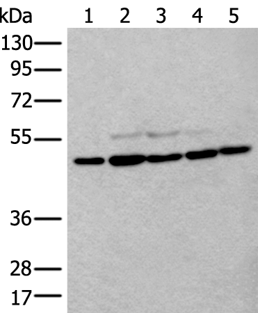
Gel: 8%SDS-PAGE Lysate: 40 microg Lane 1-5: NIH/3T3 HEPG2 Hela and 293T cell Mouse brain tissue lysates Primary antibody: TA364991 (TUBB Antibody) at dilution 1/300 Secondary antibody: Goat anti rabbit IgG at 1/8000 dilution Exposure time: 10 seconds
Gel: 8%SDS-PAGE Lysate: 40 microg Lane 1-5: NIH/3T3 HEPG2 Hela and 293T cell Mouse brain tissue lysates Primary antibody: TA364991 (TUBB Antibody) at dilution 1/300 Secondary antibody: Goat anti rabbit IgG at 1/8000 dilution Exposure time: 10 seconds
beta V Tubulin (TUBB) Rabbit Polyclonal Antibody
TA364991
Overview
- SupplierOriGene
- Product Namebeta V Tubulin (TUBB) Rabbit Polyclonal Antibody
- Delivery Days Customer14
- ApplicationsWestern Blot, ImmunoHistoChemistry
- CertificationResearch Use Only
- ClonalityPolyclonal
- Gene ID203068
- Target nameTUBB
- Target descriptiontubulin beta class I
- Target synonymsbeta Ib tubulin; CDCBM6; CSCSC1; epididymis secretory sperm binding protein; M40; OK/SW-cl.56; TUBB1; TUBB5; tubulin beta chain; tubulin beta-1 chain; tubulin beta-5 chain; tubulin, beta polypeptide
- HostRabbit
- IsotypeIgG
- Protein IDP07437
- Protein NameTubulin beta chain
- Scientific DescriptionTUBB rabbit polyclonal antibody
- Storage Instruction-20°C
- UNSPSC12352203
Related products
Product group Antibodies
ApplicationsELISA
TargetTUBB
- SizePrice

Product group Antibodies
References
beta Tubulin Polyclonal AntibodyBS-0210R
ApplicationsFlow Cytometry, ImmunoFluorescence, Western Blot, ELISA, ImmunoCytoChemistry, ImmunoHistoChemistry, ImmunoHistoChemistry Frozen, ImmunoHistoChemistry Paraffin
TargetTUBB
- SizePrice
Product group Antibodies
Anti-TUBB AntibodyA44498
ApplicationsWestern Blot, ImmunoHistoChemistry
- SizePrice
Product group Antibodies
TUBB Monoclonal AntibodyCAC12975
ApplicationsFlow Cytometry, ImmunoFluorescence, ImmunoPrecipitation, Western Blot, ELISA, ImmunoHistoChemistry
- SizePrice
Product group Antibodies

TUBB / Beta Tubulin Antibody (Internal)LS-C354310
ApplicationsWestern Blot
TargetTUBB
- SizePrice
Product group Antibodies
TUBB Monoclonal AntibodyCSB-MA080255
ApplicationsWestern Blot, ELISA, ImmunoHistoChemistry
ReactivityCanine, Chicken, Hamster, Human, Insect, Monkey, Mouse, Rabbit, Rat, Sheep, Yeast
TargetTUBB
- SizePrice

Product group Antibodies
Anti-TUBB AntibodyHPA046280
ApplicationsWestern Blot, ImmunoHistoChemistry
ReactivityHuman, Mouse, Rat
TargetTUBB
- SizePrice

Product group Antibodies
Anti-Beta Tubulin/TUBB Antibody Picoband(r)A01857-1-CARRIER-FREE
ApplicationsFlow Cytometry, ImmunoFluorescence, Western Blot, ImmunoCytoChemistry, ImmunoHistoChemistry
TargetTUBB
- SizePrice