


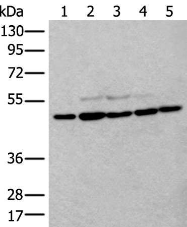
Figure 1. Western blot analysis of Beta Tubulin using anti-Beta Tubulin antibody (A01857-1). Electrophoresis was performed on a 5-20% SDS-PAGE gel at 70V (Stacking gel) / 90V (Resolving gel) for 2-3 hours. The sample well of each lane was loaded with 30 ug of sample under reducing conditions. Lane 1: human SiHa whole cell lysates, Lane 2: human 293T whole cell lysates, Lane 3: human HepG2 whole cell lysates, Lane 4: monkey COS-7 whole cell lysates, Lane 5: chicken heart tissue lysates, Lane 6: rat brain tissue lysates, Lane 7: rat PC-12 whole cell lysates, Lane 8: mouse brain tissue lysates, Lane 9: mouse NIH/3T3 whole cell lysates. After electrophoresis, proteins were transferred to a nitrocellulose membrane at 150 mA for 50-90 minutes. Blocked the membrane with 5% non-fat milk/TBS for 1.5 hour at RT. The membrane was incubated with rabbit anti-Beta Tubulin antigen affinity purified polyclonal antibody (Catalog # A01857-1) at 0.5 microg/mL overnight at 4°C, then washed with TBS-0.1%Tween 3 times with 5 minutes each and probed with a goat anti-rabbit IgG-HRP secondary antibody at a dilution of 1:5000 for 1.5 hour at RT. The signal is developed using an Enhanced Chemiluminescent detection (ECL) kit (Catalog # EK1002) with Tanon 5200 system. A specific band was detected for Beta Tubulin at approximately 50 kDa. The expected band size for Beta Tubulin is at 50 kDa.
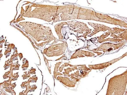
. Beta Tubulin was detected in a paraffin-embedded section of human liver cancer tissue. Heat mediated antigen retrieval was performed in EDTA buffer (pH 8.0, epitope retrieval solution). The tissue section was blocked with 10% goat serum. The tissue section was then incubated with 2 microg/ml rabbit anti-Beta Tubulin Antibody (A01857-1) overnight at 4°C. Peroxidase Conjugated Goat Anti-rabbit IgG was used as secondary antibody and incubated for 30 minutes at 37°C. The tissue section was developed using HRP Conjugated Rabbit IgG Super Vision Assay Kit (Catalog # SV0002) with DAB as the chromogen.")
. Beta Tubulin was detected in a paraffin-embedded section of human testicular germ cell tumor tissue. Heat mediated antigen retrieval was performed in EDTA buffer (pH 8.0, epitope retrieval solution). The tissue section was blocked with 10% goat serum. The tissue section was then incubated with 2 microg/ml rabbit anti-Beta Tubulin Antibody (A01857-1) overnight at 4°C. Peroxidase Conjugated Goat Anti-rabbit IgG was used as secondary antibody and incubated for 30 minutes at 37°C. The tissue section was developed using HRP Conjugated Rabbit IgG Super Vision Assay Kit (Catalog # SV0002) with DAB as the chromogen.")
. Beta Tubulin was detected in an immunocytochemical section of U20S cells. Enzyme antigen retrieval was performed using IHC enzyme antigen retrieval reagent (AR0022) for 15 mins. The cells were blocked with 10% goat serum. And then incubated with 5 microg/mL rabbit anti-Beta Tubulin Antibody (A01857-1) overnight at 4°C. Cy3 Conjugated Goat Anti-Rabbit IgG (BA1032) was used as secondary antibody at 1:100 dilution and incubated for 30 minutes at 37°C. The section was counterstained with DAPI. Visualize using a fluorescence microscope and filter sets appropriate for the label used.")
. Overlay histogram showing SiHa cells stained with A01857-1 (Blue line). To facilitate intracellular staining, cells were fixed with 4% paraformaldehyde and permeabilized with permeabilization buffer. The cells were blocked with 10% normal goat serum. And then incubated with rabbit anti-Beta Tubulin Antibody (A01857-1, 1 microg/1x106 cells) for 30 min at 20°C. DyLight®488 conjugated goat anti-rabbit IgG (BA1127, 5-10 microg/1x106 cells) was used as secondary antibody for 30 minutes at 20°C. Isotype control antibody (Green line) was rabbit IgG (1 microg/1x106) used under the same conditions. Unlabelled sample (Red line) was also used as a control.")
Figure 1. Western blot analysis of Beta Tubulin using anti-Beta Tubulin antibody (A01857-1). Electrophoresis was performed on a 5-20% SDS-PAGE gel at 70V (Stacking gel) / 90V (Resolving gel) for 2-3 hours. The sample well of each lane was loaded with 30 ug of sample under reducing conditions. Lane 1: human SiHa whole cell lysates, Lane 2: human 293T whole cell lysates, Lane 3: human HepG2 whole cell lysates, Lane 4: monkey COS-7 whole cell lysates, Lane 5: chicken heart tissue lysates, Lane 6: rat brain tissue lysates, Lane 7: rat PC-12 whole cell lysates, Lane 8: mouse brain tissue lysates, Lane 9: mouse NIH/3T3 whole cell lysates. After electrophoresis, proteins were transferred to a nitrocellulose membrane at 150 mA for 50-90 minutes. Blocked the membrane with 5% non-fat milk/TBS for 1.5 hour at RT. The membrane was incubated with rabbit anti-Beta Tubulin antigen affinity purified polyclonal antibody (Catalog # A01857-1) at 0.5 microg/mL overnight at 4°C, then washed with TBS-0.1%Tween 3 times with 5 minutes each and probed with a goat anti-rabbit IgG-HRP secondary antibody at a dilution of 1:5000 for 1.5 hour at RT. The signal is developed using an Enhanced Chemiluminescent detection (ECL) kit (Catalog # EK1002) with Tanon 5200 system. A specific band was detected for Beta Tubulin at approximately 50 kDa. The expected band size for Beta Tubulin is at 50 kDa.
Anti-Beta Tubulin/TUBB Antibody Picoband(r)
A01857-1-CARRIER-FREE
ApplicationsFlow Cytometry, ImmunoFluorescence, Western Blot, ImmunoCytoChemistry, ImmunoHistoChemistry
Product group Antibodies
ReactivityChicken, Human, Monkey, Mouse, Rat
TargetTUBB
Overview
- SupplierBoster Bio
- Product NameAnti-Beta Tubulin/TUBB Antibody Picoband(r)
- Delivery Days Customer9
- ApplicationsFlow Cytometry, ImmunoFluorescence, Western Blot, ImmunoCytoChemistry, ImmunoHistoChemistry
- CertificationResearch Use Only
- ClonalityPolyclonal
- Concentration500 ug/ml
- Gene ID203068
- Target nameTUBB
- Target descriptiontubulin beta class I
- Target synonymsCDCBM6, CSCSC1, M40, OK/SW-cl.56, TUBB1, TUBB5, tubulin beta chain, beta Ib tubulin, epididymis secretory sperm binding protein, tubulin beta-1 chain, tubulin beta-5 chain, tubulin, beta polypeptide
- HostRabbit
- IsotypeIgG
- Protein IDP07437
- Protein NameTubulin beta chain
- Scientific DescriptionBoster Bio Anti-Beta Tubulin/TUBB Antibody Picoband® catalog # A01857-1. Tested in Flow Cytometry, IF, IHC, ICC, WB applications. This antibody reacts with Human, Monkey, Mouse, Rat, Chicken. The brand Picoband indicates this is a premium antibody that guarantees superior quality, high affinity, and strong signals with minimal background in Western blot applications. Only our best-performing antibodies are designated as Picoband, ensuring unmatched performance.
- ReactivityChicken, Human, Monkey, Mouse, Rat
- Storage Instruction-20°C,2°C to 8°C
- UNSPSC12352203
Related products
Product group Antibodies
Anti-TUBB AntibodyA44498
ApplicationsWestern Blot, ImmunoHistoChemistry
ReactivityHuman, Mouse
- SizePrice
Product group Antibodies

Anti-Beta Tubulin type VI [AA16 (6A10)]Ab02684-10.0
ApplicationsImmunoFluorescence, Western Blot
ReactivityHuman, Mammals
TargetTUBB
- SizePrice
Product group Antibodies
ApplicationsELISA
ReactivityHuman
TargetTUBB
- SizePrice

Product group Antibodies
References
beta Tubulin Polyclonal AntibodyBS-0210R
ApplicationsFlow Cytometry, ImmunoFluorescence, Western Blot, ELISA, ImmunoCytoChemistry, ImmunoHistoChemistry, ImmunoHistoChemistry Frozen, ImmunoHistoChemistry Paraffin
ReactivityBovine, Human, Mouse, Porcine, Rat
TargetTUBB
- SizePrice
Product group Antibodies
TUBB Monoclonal AntibodyCSB-MA080255
ApplicationsWestern Blot, ELISA, ImmunoHistoChemistry
ReactivityCanine, Chicken, Hamster, Human, Insect, Monkey, Mouse, Rabbit, Rat, Sheep, Yeast
TargetTUBB
- SizePrice
Product group Antibodies
TUBB Monoclonal AntibodyCAC12975
ApplicationsFlow Cytometry, ImmunoFluorescence, ImmunoPrecipitation, Western Blot, ELISA, ImmunoHistoChemistry
ReactivityMouse, Rabbit, Rat
- SizePrice
Product group Antibodies
beta Tubulin antibodyGTX101279
ApplicationsImmunoFluorescence, Western Blot, ImmunoCytoChemistry, ImmunoHistoChemistry, ImmunoHistoChemistry Paraffin
ReactivityHamster, Human, Mouse, Rat, Zebra Fish
TargetTUBB
- SizePrice

Product group Antibodies
Anti-TUBB AntibodyHPA046280
ApplicationsWestern Blot, ImmunoHistoChemistry
ReactivityHuman, Mouse, Rat
TargetTUBB
- SizePrice