

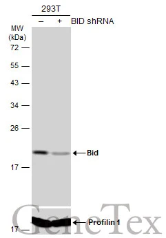
Non-transfected (–) and transfected (+) 293T whole cell extracts (60 μg) were separated by 12% SDS-PAGE, and the membrane was blotted with Bid antibody [N1C3-2] (GTX110568) diluted at 1:1000. The HRP-conjugated anti-rabbit IgG antibody (GTX213110-01) was used to detect the primary antibody.
![Bid antibody [N1C3-2] detects Bid protein by western blot analysis. A. 30 μg 293T whole cell lysate/extract B. 30 μg A431 whole cell lysate/extract C. 30 μg H1299 whole cell lysate/extract D. 30 μg HeLa whole cell lysate/extract E. 30 μg HepG2 whole cell lysate/extract F. 30 μg Molt-4 whole cell lysate/extract G. 30 μg Raji whole cell lysate/extract 7.5 % SDS-PAGE Bid antibody [N1C3-2] (GTX110568) dilution: 1:1000](https://www.genetex.com/upload/website/prouct_img/normal/GTX110568/GTX110568_40051_WB_w_23060500_928.webp "Bid antibody [N1C3-2] detects Bid protein by western blot analysis. A. 30 μg 293T whole cell lysate/extract B. 30 μg A431 whole cell lysate/extract C. 30 μg H1299 whole cell lysate/extract D. 30 μg HeLa whole cell lysate/extract E. 30 μg HepG2 whole cell lysate/extract F. 30 μg Molt-4 whole cell lysate/extract G. 30 μg Raji whole cell lysate/extract 7.5 % SDS-PAGE Bid antibody [N1C3-2] (GTX110568) dilution: 1:1000")
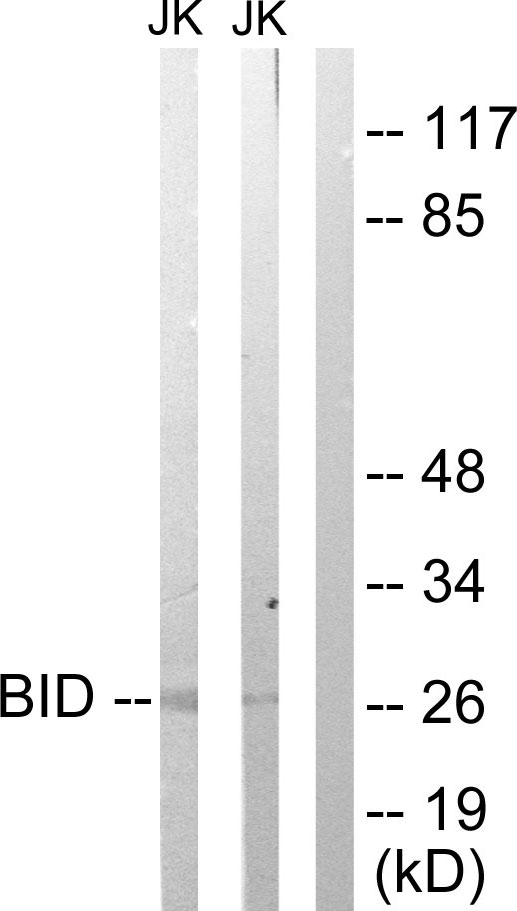
 antibody at 1:500 dilution.
Antigen Retrieval: Citrate buffer, pH 6.0, 15 min")
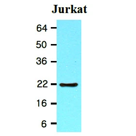
![Immunoprecipitation of Bid protein from Jurkat whole cell extracts using 5 μg of Bid antibody [N1C3-2] (GTX110568) or Bid antibody [N1C3] (GTX101323). Western blot analysis was performed using Bid antibody [N1C3-2] (GTX110568) diluted at 1:500. EasyBlot anti-Rabbit IgG (GTX221666-01) was used as a secondary reagent.](https://www.genetex.com/upload/website/prouct_img/normal/GTX110568/GTX110568_40051_IP_2_w_23060500_313.webp "Immunoprecipitation of Bid protein from Jurkat whole cell extracts using 5 μg of Bid antibody [N1C3-2] (GTX110568) or Bid antibody [N1C3] (GTX101323). Western blot analysis was performed using Bid antibody [N1C3-2] (GTX110568) diluted at 1:500. EasyBlot anti-Rabbit IgG (GTX221666-01) was used as a secondary reagent.")
![Bid antibody [N1C3-2] detects Bid protein at cytoplasm by immunofluorescent analysis. Sample: A431 cells were fixed in 4% paraformaldehyde at RT for 15 min. Green: Bid protein stained by Bid antibody [N1C3-2] (GTX110568) diluted at 1:1000. Red: alpha Tubulin, a cytoskeleton marker, stained by alpha Tubulin antibody [GT114] (GTX628802) diluted at 1:1000. Blue: Hoechst 33342 staining.](https://www.genetex.com/upload/website/prouct_img/normal/GTX110568/GTX110568_40051_20150410_IFA_w_23060500_431.webp "Bid antibody [N1C3-2] detects Bid protein at cytoplasm by immunofluorescent analysis. Sample: A431 cells were fixed in 4% paraformaldehyde at RT for 15 min. Green: Bid protein stained by Bid antibody [N1C3-2] (GTX110568) diluted at 1:1000. Red: alpha Tubulin, a cytoskeleton marker, stained by alpha Tubulin antibody [GT114] (GTX628802) diluted at 1:1000. Blue: Hoechst 33342 staining.")
![Bid antibody [N1C3-2] detects BID protein at cytoplasm by immunofluorescent analysis. Sample: HeLa cells were fixed in 2% paraformaldehyde/culture medium at 37oC for 30 min. Green: BID protein stained by Bid antibody [N1C3-2] (GTX110568) diluted at 1:500. Blue: Hoechst 33343 staining.](https://www.genetex.com/upload/website/prouct_img/normal/GTX110568/GTX110568_40051_IFA_w_23060500_209.webp "Bid antibody [N1C3-2] detects BID protein at cytoplasm by immunofluorescent analysis. Sample: HeLa cells were fixed in 2% paraformaldehyde/culture medium at 37oC for 30 min. Green: BID protein stained by Bid antibody [N1C3-2] (GTX110568) diluted at 1:500. Blue: Hoechst 33343 staining.")
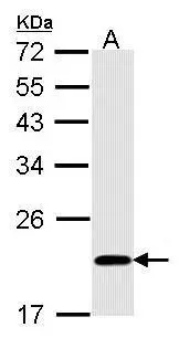
![Whole cell extract (30 μg) was separated by 12% SDS-PAGE, and the membrane was blotted with Bid antibody [N1C3-2] (GTX110568) diluted at 1:1000.](https://www.genetex.com/upload/website/prouct_img/normal/GTX110568/GTX110568_40051_20151119_WB_R_w_23060500_800.webp "Whole cell extract (30 μg) was separated by 12% SDS-PAGE, and the membrane was blotted with Bid antibody [N1C3-2] (GTX110568) diluted at 1:1000.")
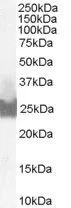
![Various whole cell extracts (30 μg) were separated by 12% SDS-PAGE, and the membrane was blotted with Bid antibody [N1C3-2] (GTX110568) diluted at 1:500.](https://www.genetex.com/upload/website/prouct_img/normal/GTX110568/GTX110568_40051_20151203_WB_M_w_23060500_187.webp "Various whole cell extracts (30 μg) were separated by 12% SDS-PAGE, and the membrane was blotted with Bid antibody [N1C3-2] (GTX110568) diluted at 1:500.")

Non-transfected (–) and transfected (+) 293T whole cell extracts (60 μg) were separated by 12% SDS-PAGE, and the membrane was blotted with Bid antibody [N1C3-2] (GTX110568) diluted at 1:1000. The HRP-conjugated anti-rabbit IgG antibody (GTX213110-01) was used to detect the primary antibody.
Bid antibody [N1C3-2]
GTX110568
ApplicationsImmunoFluorescence, ImmunoPrecipitation, Western Blot, ImmunoCytoChemistry, ImmunoHistoChemistry, ImmunoHistoChemistry Paraffin
Product group Antibodies
ReactivityHuman, Mouse, Rat
TargetBID
Overview
- SupplierGeneTex
- Product NameBid antibody [N1C3-2]
- Delivery Days Customer9
- Application Supplier NoteWB: 1:500-1:3000. ICC/IF: 1:100-1:1000. IHC-P: 1:100-1:1000. IP: 1:100-1:500. *Optimal dilutions/concentrations should be determined by the researcher.Not tested in other applications.
- ApplicationsImmunoFluorescence, ImmunoPrecipitation, Western Blot, ImmunoCytoChemistry, ImmunoHistoChemistry, ImmunoHistoChemistry Paraffin
- CertificationResearch Use Only
- ClonalityPolyclonal
- Concentration1 mg/ml
- ConjugateUnconjugated
- Gene ID637
- Target nameBID
- Target descriptionBH3 interacting domain death agonist
- Target synonymsFP497, BH3-interacting domain death agonist, Human BID coding sequence, apoptic death agonist, desmocollin type 4, p22 BID
- HostRabbit
- IsotypeIgG
- Protein IDP55957
- Protein NameBH3-interacting domain death agonist
- Scientific DescriptionThis gene encodes a death agonist that heterodimerizes with either agonist BAX or antagonist BCL2. The encoded protein is a member of the BCL-2 family of cell death regulators. It is a mediator of mitochondrial damage induced by caspase-8 (CASP8); CASP8 cleaves this encoded protein, and the COOH-terminal part translocates to mitochondria where it triggers cytochrome c release. Multiple alternatively spliced transcript variants have been found, but the full-length nature of some variants has not been defined. [provided by RefSeq]
- ReactivityHuman, Mouse, Rat
- Storage Instruction-20°C or -80°C,2°C to 8°C
- UNSPSC12352203
References
- Su CC, Yu CC, Shih YW, et al. Protective Effect of Alpha-Linolenic Acid on Human Oral Squamous Cell Carcinoma Metastasis and Apoptotic Cell Death. Nutrients. 2023,15(23). doi: 10.3390/nu15234992Read this paper
- Sikorski K, Mehta A, Inngjerdingen M, et al. A high-throughput pipeline for validation of antibodies. Nat Methods. 2018,15(11):909-912. doi: 10.1038/s41592-018-0179-8Read this paper
- Ovadje P, Ammar S, Guerrero JA, et al. Dandelion root extract affects colorectal cancer proliferation and survival through the activation of multiple death signalling pathways. Oncotarget. 2016,7(45):73080-73100. doi: 10.18632/oncotarget.11485Read this paper
- Carpentieri A, Cozzoli E, Scimeca M, et al. Differentiation of human neuroblastoma cells toward the osteogenic lineage by mTOR inhibitor. Cell Death Dis. 2015,6(11):e1974. doi: 10.1038/cddis.2015.244Read this paper
- Pu YF, Wang L, Wu HH, et al. Generation of homologous cell pairs using the oral lymphatic system. Int J Clin Exp Pathol. 2014,7(4):1563-71.Read this paper
Datasheet
Related products
Product group Antibodies
Anti-BID Antibody, Biotinylated130-10027B-50
ApplicationsELISA
ReactivityHuman
TargetBID
- SizePrice
Product group Antibodies
References
Bid antibody [N1C3]GTX101323
ApplicationsImmunoPrecipitation, Western Blot
ReactivityHuman
TargetBID
- SizePrice
Product group Antibodies
Bid antibody [4D3]GTX57542
ApplicationsImmunoFluorescence, Western Blot, ImmunoCytoChemistry
ReactivityHuman
TargetBID
- SizePrice
![ICC/IF analysis of HeLa cells using GTX60429 Bid antibody [3C5]. Green : Bid Blue: DRAQ5 fluorescent DNA dye Red: Actin filaments](https://www.genetex.com/upload/website/prouct_img/normal/GTX60429/GTX60429_20170912_ICCIF_w_23061123_825.webp)
Product group Antibodies
References
Bid antibody [3C5]GTX60429
ApplicationsFlow Cytometry, ImmunoFluorescence, Western Blot, ELISA, ImmunoCytoChemistry, ImmunoHistoChemistry, ImmunoHistoChemistry Paraffin
ReactivityHuman
TargetBID
- SizePrice
Product group Antibodies
Bid Polyclonal AntibodyCAC11767
ApplicationsELISA, ImmunoHistoChemistry
TargetBID
- SizePrice
Product group Antibodies
Bid Recombinant Antibody, AbBy Fluor-488 ConjugatedBSM-61450R-BF488
ApplicationsFlow Cytometry, ImmunoFluorescence, Western Blot, ImmunoCytoChemistry
ReactivityHuman
TargetBID
- SizePrice
Product group Antibodies
Bid antibody, C-termGTX89562
ApplicationsWestern Blot
ReactivityHuman
TargetBID
- SizePrice
Product group Antibodies
Anti-BID AntibodyA98541
ApplicationsWestern Blot, ELISA, ImmunoHistoChemistry
ReactivityHuman, Mouse
- SizePrice