

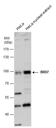
BRD7 antibody detects BRD7 protein by western blot analysis. HeLa whole cell extracts and nuclear extracts (30 μg) were separated by 7.5% SDS-PAGE, and the membrane was blotted with BRD7 antibody (GTX118755) diluted at 1:500.
![BRD7 antibody [C1C3] detects BRD7 protein at nucleus by immunofluorescent analysis. Sample: HeLa cells were fixed in 4% paraformaldehyde at RT for 15 min. Green: BRD7 protein stained by BRD7 antibody [C1C3] (GTX118755) diluted at 1:500. Red: phalloidin, a cytoskeleton marker, stained by () diluted at 1:200. Blue: Hoechst 33342 staining. Scale bar = 10 μm.](https://www.genetex.com/upload/website/prouct_img/normal/GTX118755/GTX118755_40702_20160106_IFA_w_23060519_210.webp "BRD7 antibody [C1C3] detects BRD7 protein at nucleus by immunofluorescent analysis. Sample: HeLa cells were fixed in 4% paraformaldehyde at RT for 15 min. Green: BRD7 protein stained by BRD7 antibody [C1C3] (GTX118755) diluted at 1:500. Red: phalloidin, a cytoskeleton marker, stained by () diluted at 1:200. Blue: Hoechst 33342 staining. Scale bar = 10 μm.")
BRD7 antibody detects BRD7 protein by western blot analysis. HeLa whole cell extracts and nuclear extracts (30 μg) were separated by 7.5% SDS-PAGE, and the membrane was blotted with BRD7 antibody (GTX118755) diluted at 1:500.
BRD7 antibody [C1C3]
GTX118755
ApplicationsImmunoFluorescence, Western Blot, ImmunoCytoChemistry
Product group Antibodies
ReactivityHuman
TargetBRD7
Overview
- SupplierGeneTex
- Product NameBRD7 antibody [C1C3]
- Delivery Days Customer9
- Application Supplier NoteWB: 1:500-1:3000. ICC/IF: 1:100-1:1000. *Optimal dilutions/concentrations should be determined by the researcher.Not tested in other applications.
- ApplicationsImmunoFluorescence, Western Blot, ImmunoCytoChemistry
- CertificationResearch Use Only
- ClonalityPolyclonal
- Concentration1 mg/ml
- ConjugateUnconjugated
- Gene ID29117
- Target nameBRD7
- Target descriptionbromodomain containing 7
- Target synonymsBP75, CELTIX1, NAG4, SMARCI1, bromodomain-containing protein 7, 75 kDa bromodomain protein, protein CELTIX-1
- HostRabbit
- IsotypeIgG
- Protein IDQ9NPI1
- Protein NameBromodomain-containing protein 7
- Scientific DescriptionThis gene encodes a protein which is a member of the bromodomain-containing protein family. The product of this gene has been identified as a component of one form of the SWI/SNF chromatin remodeling complex, and as a protein which interacts with p53 and is required for p53-dependent oncogene-induced senescence which prevents tumor growth. Pseudogenes have been described on chromosomes 2, 3, 6, 13 and 14. Alternative splicing results in multiple transcript variants. [provided by RefSeq]
- ReactivityHuman
- Storage Instruction-20°C or -80°C,2°C to 8°C
- UNSPSC41116161
Datasheet
Related products
Product group Antibodies
Anti-BRD7 Antibody Picoband(r)A01289-1-CARRIER-FREE
ApplicationsWestern Blot, ELISA, ImmunoHistoChemistry
ReactivityHuman, Mouse, Rat
TargetBRD7
- SizePrice
Product group Antibodies

Anti-BRD7 [RAB-C282]Ab01745-1.1
ApplicationsImmunoFluorescence, ImmunoPrecipitation
ReactivityHuman
TargetBRD7
- SizePrice
Product group Antibodies

BRD7 AntibodyLS-C831914
ApplicationsELISA, ImmunoHistoChemistry
ReactivityHuman, Mouse
TargetBRD7
- SizePrice

Product group Antibodies
Anti-BRD7 AntibodyHPA060171
ApplicationsImmunoCytoChemistry
ReactivityHuman
TargetBRD7
- SizePrice
Product group Antibodies
BRD7 AntibodyCSB-PA865097LA01HU
ApplicationsImmunoFluorescence, ELISA, ImmunoHistoChemistry
ReactivityHuman
TargetBRD7
- SizePrice
Product group Antibodies
Anti-BRD7 Antibody144-02308
ApplicationsWestern Blot
ReactivityHuman
TargetBRD7
- SizePrice
Product group Antibodies
BRD7 Polyclonal AntibodyBS-7779R
ApplicationsImmunoFluorescence, ELISA, ImmunoCytoChemistry, ImmunoHistoChemistry, ImmunoHistoChemistry Frozen, ImmunoHistoChemistry Paraffin
ReactivityBovine, Canine, Human, Mouse, Porcine, Rabbit, Rat, Sheep
TargetBRD7
- SizePrice