

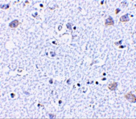
IHC-P analysis of human brain tissue using GTX85054 BRSK1 antibody. Working concentration : 5 μg/ml
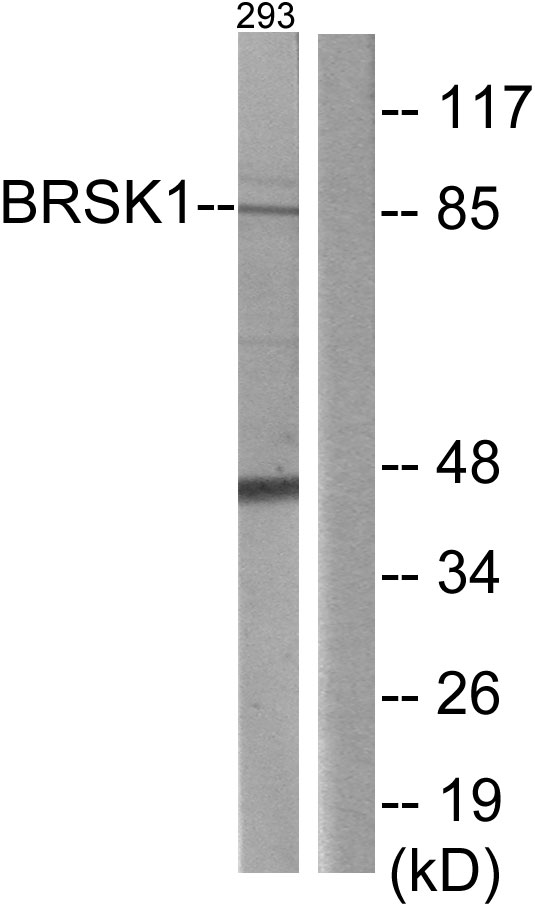
 0.5, (B) 1, and (C) 2 μg/ml")

IHC-P analysis of human brain tissue using GTX85054 BRSK1 antibody. Working concentration : 5 μg/ml
BRSK1 antibody
GTX85054
ApplicationsWestern Blot, ELISA, ImmunoHistoChemistry, ImmunoHistoChemistry Paraffin
Product group Antibodies
ReactivityHuman, Mouse, Rat
TargetBRSK1
Overview
- SupplierGeneTex
- Product NameBRSK1 antibody
- Delivery Days Customer9
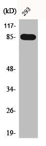
- Application Supplier NoteWB: 0.5 - 2 microg/mL. IHC-P: 5 microg/mL. *Optimal dilutions/concentrations should be determined by the researcher.Not tested in other applications.
- ApplicationsWestern Blot, ELISA, ImmunoHistoChemistry, ImmunoHistoChemistry Paraffin
- CertificationResearch Use Only
- ClonalityPolyclonal
- Concentration1 mg/ml
- ConjugateUnconjugated
- Gene ID84446
- Target nameBRSK1
- Target descriptionBR serine/threonine kinase 1
- Target synonymsSAD-B, hSAD1, serine/threonine-protein kinase BRSK1, BR serine/threonine-protein kinase 1, SAD1 homolog, SAD1 kinase, brain-selective kinase 1, brain-specific serine/threonine-protein kinase 1, protein kinase SAD1A, serine/threonine-protein kinase SAD-B, synapses of Amphids Defective homolog 1
- HostRabbit
- IsotypeIgG
- Protein IDQ8TDC3
- Protein NameSerine/threonine-protein kinase BRSK1
- Scientific DescriptionBRSK1 was initially identified as a mammalian homolog to the fission yeast S. pombe Cdr2, a mitosis-regulatory kinase and also shows significant homology to the C. elegans neuronal cell polarity regulator SAD1. BRSK1 is unbiquitously expressed, with highest levels of expression in the brain and testes. Similar to its yeast homolog, BRSK1 is thought to be involved in stress-induced cell cycle arrest. Overexpression of this protein leads to the G2/M arrest in HeLa S2 cells and UV-induced G2/M arrest could be partially abrogated by reduced expression of BRSK1 through the use of siRNA, indicating its role in DNA damage checkpoint function. More recently, it has been shown that both BRSK1 and the related protein BRSK2 are required for mammalian neuronal polarization. While BRSK1- and BRSK2- mice were viable, double-mutant mice died within two hours of birth. Neurons from these mice showed uniformly-sized neurites as opposed to the normal long axon and multiple shorter dendrites. These neurites also displayed both axonal and dendritic markers. At least two isoforms of BRSK1 are known to exist.
- ReactivityHuman, Mouse, Rat
- Storage Instruction-20°C or -80°C,2°C to 8°C
- UNSPSC12352203
Datasheet
Related products
Product group Antibodies
Anti-Mouse BRSK1 (Center) Antibody102-23694
ApplicationsWestern Blot
TargetBRSK1
- SizePrice
Product group Antibodies
BRSK1 Polyclonal AntibodyCAC14697
ApplicationsWestern Blot, ELISA, ImmunoHistoChemistry
ReactivityMouse
TargetBRSK1
- SizePrice
Product group Antibodies
BRSK1 Polyclonal AntibodyBS-7905R
ApplicationsImmunoFluorescence, ELISA, ImmunoCytoChemistry, ImmunoHistoChemistry, ImmunoHistoChemistry Frozen, ImmunoHistoChemistry Paraffin
ReactivityBovine, Equine, Human, Mouse, Porcine, Rat
TargetBRSK1
- SizePrice
Product group Antibodies
BRSK1 AntibodyCSB-PA001058
ApplicationsImmunoFluorescence, Western Blot, ELISA
ReactivityHuman, Mouse
TargetBRSK1
- SizePrice
Product group Antibodies
Anti-BRSK1 AntibodyA99163
ApplicationsWestern Blot, ELISA
ReactivityHuman, Mouse
- SizePrice
Product group Antibodies

KIAA1811 / BRSK1 AntibodyLS-C831919
ApplicationsELISA, ImmunoHistoChemistry
ReactivityHuman, Mouse, Rat
TargetBRSK1
- SizePrice

Product group Antibodies
Anti-BRSK1 AntibodyHPA061719
ApplicationsWestern Blot, ImmunoHistoChemistry
ReactivityHuman
TargetBRSK1
- SizePrice
Product group Antibodies
BRSK1 antibodyGTX85055
ApplicationsImmunoFluorescence, Western Blot, ELISA, ImmunoCytoChemistry, ImmunoHistoChemistry, ImmunoHistoChemistry Paraffin
ReactivityHuman, Mouse, Rat
TargetBRSK1
- SizePrice

Product group Antibodies
Anti-BRSK1 Antibody Picoband(r)A05997-2-CARRIER-FREE
ApplicationsFlow Cytometry, ImmunoFluorescence, Western Blot, ELISA, ImmunoCytoChemistry, ImmunoHistoChemistry
ReactivityHuman, Mouse, Rat
TargetBRSK1
- SizePrice