

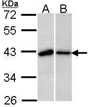
Sample (30 ug of whole cell lysate) A: 293T B: H1299 10% SDS PAGE GTX113595 diluted at 1:1000
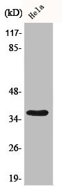
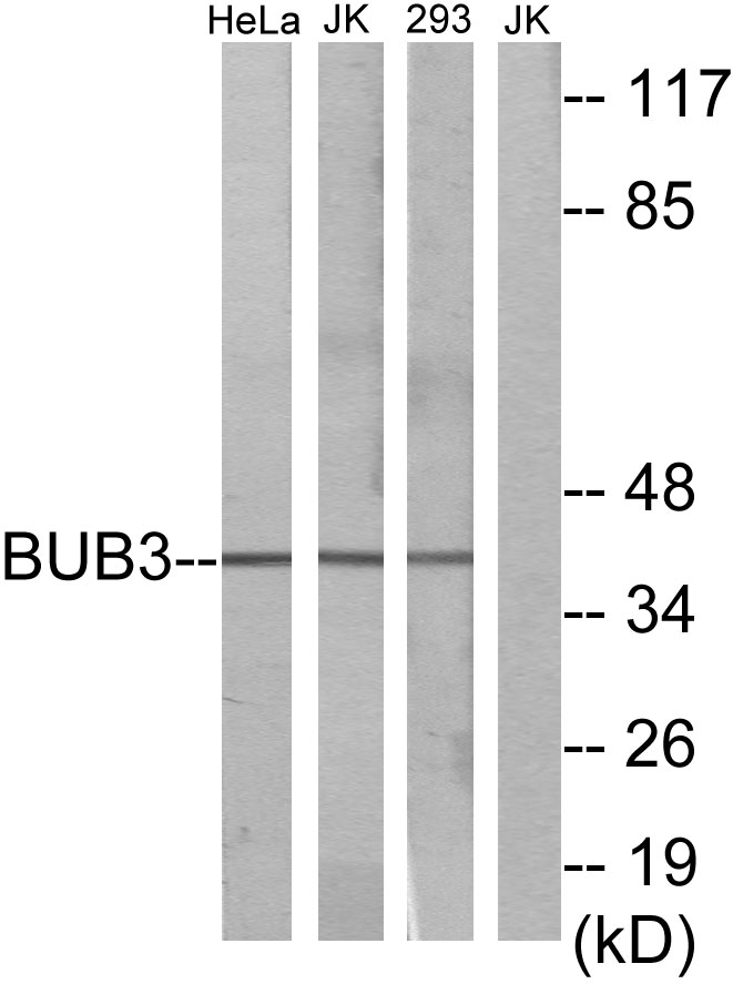
![Various whole cell extracts (30 μg) were separated by 10% SDS-PAGE, and the membrane was blotted with BUB3 antibody [N1C1] (GTX113595) diluted at 1:500. The HRP-conjugated anti-rabbit IgG antibody (GTX213110-01) was used to detect the primary antibody.](https://www.genetex.com/upload/website/prouct_img/normal/GTX113595/GTX113595_40142_20170901_WB_R_w_23060501_632.webp "Various whole cell extracts (30 μg) were separated by 10% SDS-PAGE, and the membrane was blotted with BUB3 antibody [N1C1] (GTX113595) diluted at 1:500. The HRP-conjugated anti-rabbit IgG antibody (GTX213110-01) was used to detect the primary antibody.")
 were separated by 10% SDS-PAGE, and the membrane was blotted with BUB3 antibody (GTX113595) diluted by 1:1000.")
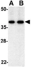
![Immunoprecipitation of BUB3 protein from 293T whole cell extracts using 5 μg of BUB3 antibody [N1C1] (GTX113595). Western blot analysis was performed using BUB3 antibody [N1C1] (GTX113595). EasyBlot anti-Rabbit IgG (GTX221666-01) was used as a secondary reagent.](https://www.genetex.com/upload/website/prouct_img/normal/GTX113595/GTX113595_40142_20150618_IP_w_23060501_447.webp "Immunoprecipitation of BUB3 protein from 293T whole cell extracts using 5 μg of BUB3 antibody [N1C1] (GTX113595). Western blot analysis was performed using BUB3 antibody [N1C1] (GTX113595). EasyBlot anti-Rabbit IgG (GTX221666-01) was used as a secondary reagent.")
 antibody at 1:100 dilution.
Antigen Retrieval: Trilogy? (EDTA based, pH 8.0) buffer, 15min")
![Various whole cell extracts (30 μg) were separated by 10% SDS-PAGE, and the membrane was blotted with BUB3 antibody [N1C1] (GTX113595) diluted at 1:500. The HRP-conjugated anti-rabbit IgG antibody (GTX213110-01) was used to detect the primary antibody.](https://www.genetex.com/upload/website/prouct_img/normal/GTX113595/GTX113595_40142_20170901_WB_M_w_23060501_363.webp "Various whole cell extracts (30 μg) were separated by 10% SDS-PAGE, and the membrane was blotted with BUB3 antibody [N1C1] (GTX113595) diluted at 1:500. The HRP-conjugated anti-rabbit IgG antibody (GTX213110-01) was used to detect the primary antibody.")
Sample (30 ug of whole cell lysate) A: 293T B: H1299 10% SDS PAGE GTX113595 diluted at 1:1000
BUB3 antibody [N1C1]
GTX113595
ApplicationsImmunoPrecipitation, Western Blot, ImmunoHistoChemistry, ImmunoHistoChemistry Paraffin
Product group Antibodies
ReactivityHuman, Mouse, Rat
TargetBUB3
Overview
- SupplierGeneTex
- Product NameBUB3 antibody [N1C1]
- Delivery Days Customer9
- Application Supplier NoteWB: 1:500-1:3000. IHC-P: 1:100-1:1000. IP: 1:100-1:500. *Optimal dilutions/concentrations should be determined by the researcher.Not tested in other applications.
- ApplicationsImmunoPrecipitation, Western Blot, ImmunoHistoChemistry, ImmunoHistoChemistry Paraffin
- CertificationResearch Use Only
- ClonalityPolyclonal
- Concentration0.32 mg/ml
- ConjugateUnconjugated
- Gene ID9184
- Target nameBUB3
- Target descriptionBUB3 mitotic checkpoint protein
- Target synonymsBUB3L, hBUB3, mitotic checkpoint protein BUB3, BUB3 budding uninhibited by benzimidazoles 3 homolog, budding uninhibited by benomyl, budding uninhibited by benzimidazoles 3 homolog, mitotic checkpoint component, testicular tissue protein Li 27
- HostRabbit
- IsotypeIgG
- Protein IDO43684
- Protein NameMitotic checkpoint protein BUB3
- Scientific DescriptionThis gene encodes a protein involved in spindle checkpoint function. The encoded protein contains four WD repeat domains and has sequence similarity with the yeast BUB3 protein. Alternate transcriptional splice variants, encoding different isoforms, have been characterized. [provided by RefSeq]
- ReactivityHuman, Mouse, Rat
- Storage Instruction-20°C or -80°C,2°C to 8°C
- UNSPSC41116161
Datasheet
Related products
Product group Antibodies
BUB3 AntibodyCSB-PA001070
ApplicationsImmunoFluorescence, Western Blot, ELISA, ImmunoHistoChemistry
ReactivityHuman, Mouse, Rat
TargetBUB3
- SizePrice

Product group Antibodies
Anti-BUB3 Antibody Picoband(r)A03118-2-CARRIER-FREE
ApplicationsFlow Cytometry, Western Blot, ELISA
ReactivityHuman, Rat
TargetBUB3
- SizePrice
Product group Antibodies
Anti-BUB3 AntibodyA98261
ApplicationsWestern Blot, ELISA
ReactivityHuman, Mouse, Rat
- SizePrice

Product group Antibodies
Anti-BUB3 AntibodyHPA003601
ApplicationsWestern Blot, ImmunoCytoChemistry, ImmunoHistoChemistry
ReactivityHuman, Mouse
TargetBUB3
- SizePrice
Product group Antibodies

BUB3 AntibodyLS-C334784
ApplicationsImmunoFluorescence, Western Blot, ImmunoHistoChemistry
ReactivityHuman, Mouse, Rat
TargetBUB3
- SizePrice
Product group Antibodies
BUB3 antibodyGTX31314
ApplicationsImmunoFluorescence, Western Blot, ELISA, ImmunoCytoChemistry
ReactivityHuman, Mouse
TargetBUB3
- SizePrice
Product group Antibodies
BUB3 Polyclonal AntibodyCAC14581
ApplicationsWestern Blot, ELISA, ImmunoHistoChemistry
ReactivityMouse
TargetBUB3
- SizePrice

Product group Antibodies
BUB3 Recombinant AntibodyBSM-54413R
ApplicationsFlow Cytometry, ImmunoFluorescence, ImmunoPrecipitation, Western Blot, ImmunoCytoChemistry, ImmunoHistoChemistry, ImmunoHistoChemistry Paraffin
ReactivityHuman, Mouse, Rat
TargetBUB3
- SizePrice