

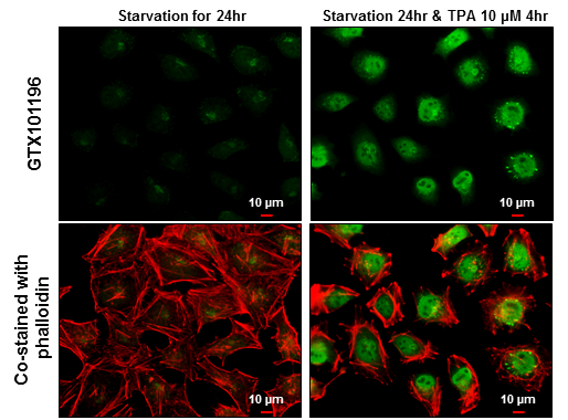
c-Fos antibody detects c-Fos protein at cytoplasm and nucleus by immunofluorescent analysis. Sample: HeLa cells were fixed in 4% paraformaldehyde at RT for 15 min. Green: c-Fos protein stained by c-Fos antibody (GTX101196) diluted at 1:500. Red: phalloidin, a cytoskeleton marker, diluted at 1:50. Scale bar = 10 μm.
![c-Fos antibody detects c-Fos protein by immunofluorescent analysis. Sample: DIV9 rat E18 primary cortical neuron cells were fixed in 4% paraformaldehyde at RT for 15 min. Green: c-Fos stained by c-Fos antibody (GTX101196) diluted at 1:1500. Red: beta Tubulin 3/ Tuj1, stained by beta Tubulin 3/ Tuj1 antibody [GT11710] (GTX631836) diluted at 1:500. Blue: Fluoroshield with DAPI (GTX30920).](https://www.genetex.com/upload/website/prouct_img/normal/GTX101196/GTX101196_40954_20181115_ICC_IF_R_w_23060100_552.webp "c-Fos antibody detects c-Fos protein by immunofluorescent analysis. Sample: DIV9 rat E18 primary cortical neuron cells were fixed in 4% paraformaldehyde at RT for 15 min. Green: c-Fos stained by c-Fos antibody (GTX101196) diluted at 1:1500. Red: beta Tubulin 3/ Tuj1, stained by beta Tubulin 3/ Tuj1 antibody [GT11710] (GTX631836) diluted at 1:500. Blue: Fluoroshield with DAPI (GTX30920).")
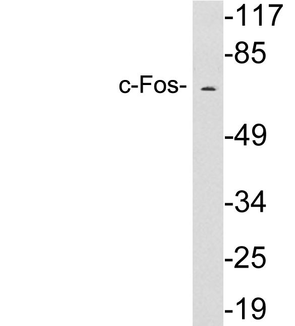
 B. 30 μg MCF-7 whole cell lysate/extract (500nM PMA treatment for 0.5hr) C. 30 μg MCF-7 whole cell lysate/extract (500nM PMA treatment for 1hr) D. 30 μg MCF-7 whole cell lysate/extract (500nM PMA treatment for 2hr) 10 % SDS-PAGE c-Fos antibody (GTX101196) dilution: 1:5000")
c-Fos antibody detects c-Fos protein at cytoplasm and nucleus by immunofluorescent analysis. Sample: HeLa cells were fixed in 4% paraformaldehyde at RT for 15 min. Green: c-Fos protein stained by c-Fos antibody (GTX101196) diluted at 1:500. Red: phalloidin, a cytoskeleton marker, diluted at 1:50. Scale bar = 10 μm.
c-Fos antibody
GTX101196
ApplicationsImmunoFluorescence, Western Blot, ImmunoCytoChemistry
Product group Antibodies
ReactivityHuman, Rat
TargetFOS
Overview
- SupplierGeneTex
- Product Namec-Fos antibody
- Delivery Days Customer9
- Application Supplier NoteWB: 1:1000-1:10000. ICC/IF: 1:100-1:1000. *Optimal dilutions/concentrations should be determined by the researcher.Not tested in other applications.
- ApplicationsImmunoFluorescence, Western Blot, ImmunoCytoChemistry
- CertificationResearch Use Only
- ClonalityPolyclonal
- Concentration1 mg/ml
- ConjugateUnconjugated
- Gene ID2353
- Target nameFOS
- Target descriptionFos proto-oncogene, AP-1 transcription factor subunit
- Target synonymsAP-1, C-FOS, p55, protein c-Fos, FBJ murine osteosarcoma viral (v-fos) oncogene homolog (oncogene FOS), FBJ murine osteosarcoma viral oncogene homolog, Fos proto-oncogene, AP-1 trancription factor subunit, G0/G1 switch regulatory protein 7, activator protein 1, cellular oncogene c-fos, proto-oncogene c-Fos, transcription factor AP-1 subunit c-Fos
- HostRabbit
- IsotypeIgG
- Protein IDP01100
- Protein NameProtein c-Fos
- Scientific DescriptionThe Fos gene family consists of 4 members: FOS, FOSB, FOSL1, and FOSL2. These genes encode leucine zipper proteins that can dimerize with proteins of the JUN family, thereby forming the transcription factor complex AP-1. As such, the FOS proteins have been implicated as regulators of cell proliferation, differentiation, and transformation. In some cases, expression of the FOS gene has also been associated with apoptotic cell death. [provided by RefSeq]
- ReactivityHuman, Rat
- Storage Instruction-20°C or -80°C,2°C to 8°C
- UNSPSC41116161
Datasheet
Related products
Product group Antibodies
Anti-c-Fos AntibodyA97643
ApplicationsWestern Blot, ELISA
ReactivityHuman, Mouse, Rat
- SizePrice
Product group Antibodies

Anti-cFos [C2-82]Ab02287-10.0
ApplicationsImmunoFluorescence, Western Blot, ELISA, ImmunoHistoChemistry
ReactivityHuman
TargetFOS
- SizePrice
Product group Antibodies
Anti-FOS Antibody144-00236
ApplicationsWestern Blot, ImmunoHistoChemistry
ReactivityHuman, Mouse
TargetFOS
- SizePrice

Product group Antibodies
Anti-FOS AntibodyAMAB91417
ApplicationsWestern Blot
ReactivityHuman
TargetFOS
- SizePrice
Product group Antibodies

FOS / c-FOS Antibody (clone AWUE3)LS-C764956
ApplicationsWestern Blot, ImmunoHistoChemistry, ImmunoHistoChemistry Paraffin
ReactivityHuman, Mouse, Rat
TargetFOS
- SizePrice

Product group Antibodies
Anti-c-Fos/FOS Antibody Picoband(r)A00297-1-CARRIER-FREE
ApplicationsFlow Cytometry, Western Blot, ELISA
ReactivityHuman
TargetFOS
- SizePrice
Product group Antibodies
References
c-fos Polyclonal Antibodybs-0469R
ApplicationsImmunoFluorescence, Western Blot, ELISA, ImmunoCytoChemistry, ImmunoHistoChemistry, ImmunoHistoChemistry Frozen, ImmunoHistoChemistry Paraffin
ReactivityBovine, Canine, Equine, Human, Mouse, Porcine, Rabbit, Rat
TargetFOS
- SizePrice
Product group Antibodies
FOS Monoclonal AntibodyCSB-MA080264
ApplicationsWestern Blot, ELISA
ReactivityHuman, Mouse, Rat
TargetFOS
- SizePrice
Product group Antibodies

Goat anti-c-FOS (aa283-295)EB11742
ApplicationsImmunoFluorescence, ELISA, ImmunoHistoChemistry
ReactivityBovine, Canine, Human, Porcine
TargetFOS
- SizePrice
Product group Antibodies
ApplicationsImmunoPrecipitation, Western Blot, ImmunoCytoChemistry, ImmunoHistoChemistry
TargetFOS
- SizePrice