

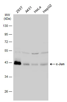
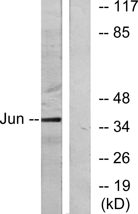
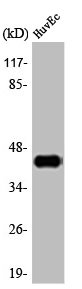
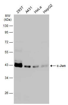
Various whole cell extracts (30 μg) were separated by 10% SDS-PAGE, and the membrane was blotted with c-Jun antibody (GTX112974) diluted at 1:1000. The HRP-conjugated anti-rabbit IgG antibody (GTX213110-01) was used to detect the primary antibody.
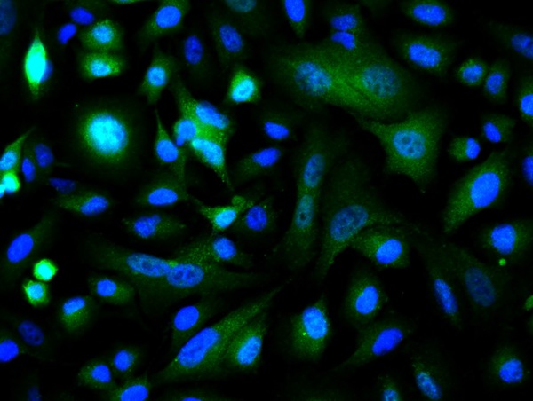
 diluted at 1:500. Red: phalloidin, a cytoskeleton marker, diluted at 1:100. Scale bar= 10 μm.")
 diluted at 1:500. Antigen Retrieval: Citrate buffer, pH 6.0, 15 min")
Various whole cell extracts (30 μg) were separated by 10% SDS-PAGE, and the membrane was blotted with c-Jun antibody (GTX112974) diluted at 1:1000. The HRP-conjugated anti-rabbit IgG antibody (GTX213110-01) was used to detect the primary antibody.
c-Jun antibody
GTX112974
ApplicationsImmunoFluorescence, Western Blot, ImmunoCytoChemistry, ImmunoHistoChemistry, ImmunoHistoChemistry Paraffin
Product group Antibodies
ReactivityHuman
TargetJUN
Overview
- SupplierGeneTex
- Product Namec-Jun antibody
- Delivery Days Customer9
- Application Supplier NoteWB: 1:500-1:3000. ICC/IF: 1:100-1:1000. *Optimal dilutions/concentrations should be determined by the researcher.Not tested in other applications.
- ApplicationsImmunoFluorescence, Western Blot, ImmunoCytoChemistry, ImmunoHistoChemistry, ImmunoHistoChemistry Paraffin
- CertificationResearch Use Only
- ClonalityPolyclonal
- Concentration0.43 mg/ml
- ConjugateUnconjugated
- Gene ID3725
- Target nameJUN
- Target descriptionJun proto-oncogene, AP-1 transcription factor subunit
- Target synonymsAP-1, AP1, c-Jun, cJUN, p39, transcription factor Jun, Jun activation domain binding protein, activator protein 1, enhancer-binding protein AP1, jun oncogene, proto-oncogene c-Jun, proto-oncogene cJun, transcription factor AP-1, transcription factor AP-1 subunit Jun, v-jun avian sarcoma virus 17 oncogene homolog, v-jun sarcoma virus 17 oncogene homolog
- HostRabbit
- IsotypeIgG
- Protein IDP05412
- Protein NameTranscription factor Jun
- Scientific DescriptionThis gene is the putative transforming gene of avian sarcoma virus 17. It encodes a protein which is highly similar to the viral protein, and which interacts directly with specific target DNA sequences to regulate gene expression. This gene is intronless and is mapped to 1p32-p31, a chromosomal region involved in both translocations and deletions in human malignancies. [provided by RefSeq, Jul 2008]
- ReactivityHuman
- Storage Instruction-20°C or -80°C,2°C to 8°C
- UNSPSC41116161
Datasheet
Related products
Product group Antibodies
Anti-c-Jun AntibodyA95242
ApplicationsImmunoPrecipitation, Western Blot, ELISA, ImmunoHistoChemistry
ReactivityHuman, Mouse, Rat
- SizePrice
Product group Antibodies
Anti-c-Jun Antibody144-61581
ApplicationsImmunoFluorescence, Western Blot
ReactivityHuman, Mouse, Rat
TargetJUN
- SizePrice

Product group Antibodies
Anti-JUN AntibodyAMAB91587
ApplicationsImmunoCytoChemistry, ImmunoHistoChemistry
ReactivityHuman
TargetJUN
- SizePrice

Product group Antibodies
Anti-c-Jun/JUN Antibody Picoband(r)A02038-2-CARRIER-FREE
ApplicationsImmunoFluorescence, Western Blot, ImmunoCytoChemistry, ImmunoHistoChemistry
ReactivityHuman
TargetJUN
- SizePrice
Product group Antibodies
References
c-Jun Polyclonal AntibodyBS-0670R
ApplicationsFlow Cytometry, ImmunoFluorescence, Western Blot, ELISA, ImmunoCytoChemistry, ImmunoHistoChemistry, ImmunoHistoChemistry Frozen, ImmunoHistoChemistry Paraffin
ReactivityBovine, Canine, Chicken, Human, Mouse, Porcine, Rat, Sheep
TargetJUN
- SizePrice
Product group Antibodies
JUN AntibodyCSB-PA000886
ApplicationsImmunoFluorescence, Western Blot, ELISA, ImmunoHistoChemistry
ReactivityHuman, Mouse, Rat
TargetJUN
- SizePrice
Product group Antibodies
Jun Polyclonal AntibodyCAC09196
ApplicationsImmunoFluorescence, Western Blot, ELISA
ReactivityMouse, Rat
TargetJUN
- SizePrice
Product group Antibodies
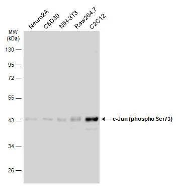
c-Jun (phospho Ser73) antibodyGTX133873
ApplicationsImmunoFluorescence, Western Blot, ImmunoCytoChemistry, ImmunoHistoChemistry, ImmunoHistoChemistry Paraffin
ReactivityHuman, Mouse, Rat
TargetJUN
- SizePrice
Product group Antibodies
c-Jun antibodyGTX134395
ApplicationsImmunoFluorescence, Western Blot, ImmunoCytoChemistry
ReactivityHuman, Mouse
TargetJUN
- SizePrice