

C-Ros Oncogene 1, Receptor Tyrosine Kinase (ROS1) Polyclonal Antibody
CAU22857
ApplicationsWestern Blot, ELISA, ImmunoCytoChemistry, ImmunoHistoChemistry, ImmunoHistoChemistry Frozen, ImmunoHistoChemistry Paraffin
Product group Antibodies
TargetROS1
Overview
- SupplierBiomatik
- Product NameC-Ros Oncogene 1, Receptor Tyrosine Kinase (ROS1) Polyclonal Antibody
- Delivery Days Customer12
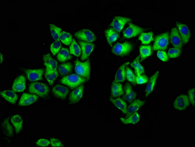
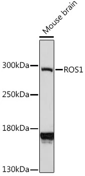
- ApplicationsWestern Blot, ELISA, ImmunoCytoChemistry, ImmunoHistoChemistry, ImmunoHistoChemistry Frozen, ImmunoHistoChemistry Paraffin
- Applications SupplierWB; ICC; IHC-P; IHC-F; ELISA.
- CertificationResearch Use Only
- ClonalityPolyclonal
- Concentration1 mg/ml
- ConjugateUnconjugated
- Gene ID6098
- Target nameROS1
- Target descriptionROS proto-oncogene 1, receptor tyrosine kinase
- Target synonymsMCF3, ROS, c-ros-1, proto-oncogene tyrosine-protein kinase ROS, ROS proto-oncogene 1 , receptor tyrosine kinase, c-ros oncogene 1 , receptor tyrosine kinase, proto-oncogene c-Ros-1, transmembrane tyrosine-specific protein kinase, v-ros avian UR2 sarcoma virus oncogene homolog 1
- HostRabbit
- Protein IDP08922
- Protein NameProto-oncogene tyrosine-protein kinase ROS
- Scientific DescriptionThe C-Ros Oncogene 1, Receptor Tyrosine Kinase (ROS1) Polyclonal Antibody (Species: Human) has been validated for the following applications: WB, ICC, IHC-P, IHC-F, ELISA.
- Reactivity SupplierHuman
- Storage Instruction-20°C,2°C to 8°C
- UNSPSC12352203
Related products
Product group Antibodies

Anti-ROS1 [ROS1-5F2]Ab03010-10.0
ApplicationsWestern Blot, ELISA
ReactivityHuman
TargetROS1
- SizePrice

Product group Antibodies
Anti-ROS1 Antibody (C-term)A04186-1
ApplicationsWestern Blot
ReactivityHuman
TargetROS1
- SizePrice

Product group Antibodies
Anti-ROS1 AntibodyA88936
ApplicationsWestern Blot
ReactivityHuman, Mouse
- SizePrice

Product group Antibodies
ROS1 Polyclonal AntibodyBS-2504R
ApplicationsFlow Cytometry, Western Blot, ELISA, ImmunoHistoChemistry, ImmunoHistoChemistry Paraffin
ReactivityHuman, Rat
TargetROS1
- SizePrice

Product group Antibodies
ROS1 AntibodyCSB-PA020072LA01HU
ApplicationsImmunoFluorescence, ELISA
ReactivityHuman
TargetROS1
- SizePrice
Product group Antibodies

ROS1 / ROS AntibodyLS-C402768
ApplicationsELISA, ImmunoHistoChemistry
ReactivityHuman
TargetROS1
- SizePrice
Product group Antibodies
ROS antibodyGTX00638
ApplicationsWestern Blot
ReactivityHuman, Mouse
TargetROS1
- SizePrice