

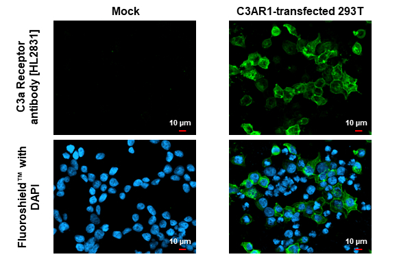
C3a Receptor antibody [HL2831] detects C3a Receptor protein by immunofluorescent analysis. Sample: Mock and transfected 293T cells were fixed in ice-cold MeOH for 5 min. Green: C3a Receptor stained by C3a Receptor antibody [HL2831] (GTX640102) diluted at 1:500. Blue: Fluoroshield with DAPI (GTX30920).
![C3a Receptor antibody [HL2831] detects C3a Receptor protein at cell membrane and cytoplasm by immunohistochemical analysis. Sample: Paraffin-embedded human glioblastoma. C3a Receptor stained by C3a Receptor antibody [HL2831] (GTX640102) diluted at 1:100. Antigen Retrieval: Citrate buffer, pH 6.0, 15 min](https://www.genetex.com/upload/website/prouct_img/normal/GTX640102/GTX640102_T-45355_20240419_IHC-P_1_24052202_523.webp "C3a Receptor antibody [HL2831] detects C3a Receptor protein at cell membrane and cytoplasm by immunohistochemical analysis. Sample: Paraffin-embedded human glioblastoma. C3a Receptor stained by C3a Receptor antibody [HL2831] (GTX640102) diluted at 1:100. Antigen Retrieval: Citrate buffer, pH 6.0, 15 min")
![C3a Receptor antibody [HL2831] detects C3a Receptor protein at cell membrane and cytoplasm by immunohistochemical analysis. Sample: Paraffin-embedded HL-60 xenograft. C3a Receptor stained by C3a Receptor antibody [HL2831] (GTX640102) diluted at 1:100. Antigen Retrieval: Citrate buffer, pH 6.0, 15 min](https://www.genetex.com/upload/website/prouct_img/normal/GTX640102/GTX640102_T-45355_20240419_IHC-P_24052202_595.webp "C3a Receptor antibody [HL2831] detects C3a Receptor protein at cell membrane and cytoplasm by immunohistochemical analysis. Sample: Paraffin-embedded HL-60 xenograft. C3a Receptor stained by C3a Receptor antibody [HL2831] (GTX640102) diluted at 1:100. Antigen Retrieval: Citrate buffer, pH 6.0, 15 min")
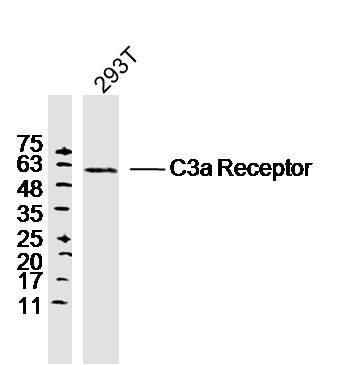
![Non-transfected (–) and transfected (+) boiled and unboiled 293T whole cell extracts (30 μg) were separated by 10% SDS-PAGE, and the membrane was blotted with C3a Receptor antibody [HL2831] (GTX640102) diluted at 1:5000. The HRP-conjugated anti-rabbit IgG antibody (GTX213110-01) was used to detect the primary antibody.](https://www.genetex.com/upload/website/prouct_img/normal/GTX640102/GTX640102_45446_20240705_WB_B_ub_24070822_172.webp "Non-transfected (–) and transfected (+) boiled and unboiled 293T whole cell extracts (30 μg) were separated by 10% SDS-PAGE, and the membrane was blotted with C3a Receptor antibody [HL2831] (GTX640102) diluted at 1:5000. The HRP-conjugated anti-rabbit IgG antibody (GTX213110-01) was used to detect the primary antibody.")
![C3a Receptor antibody [HL2831] detects C3a Receptor protein by immunohistochemical analysis. Sample: Paraffin-embedded human cerebellum. C3a Receptor stained by C3a Receptor antibody [HL2831] (GTX640102) diluted at 1:100. Antigen Retrieval: Citrate buffer, pH 6.0, 15 min](https://www.genetex.com/upload/website/prouct_img/normal/GTX640102/GTX640102_45446_20240802_IHC-P_24081300_484.webp "C3a Receptor antibody [HL2831] detects C3a Receptor protein by immunohistochemical analysis. Sample: Paraffin-embedded human cerebellum. C3a Receptor stained by C3a Receptor antibody [HL2831] (GTX640102) diluted at 1:100. Antigen Retrieval: Citrate buffer, pH 6.0, 15 min")
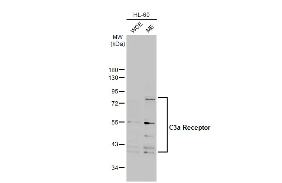
![Untreated (–) and treated (+) unboiled HL-60 whole cell extracts (30 μg) were separated by 10% SDS-PAGE, and the membrane was blotted with C3a Receptor antibody [HL2831] (GTX640102) diluted at 1:1000. The HRP-conjugated anti-rabbit IgG antibody (GTX213110-01) was used to detect the primary antibody, and the signal was developed with Trident ECL plus-Enhanced.](https://www.genetex.com/upload/website/prouct_img/normal/GTX640102/GTX640102_T-45355_20240913_WB_treatment_PNGaseF_24091901_108.webp "Untreated (–) and treated (+) unboiled HL-60 whole cell extracts (30 μg) were separated by 10% SDS-PAGE, and the membrane was blotted with C3a Receptor antibody [HL2831] (GTX640102) diluted at 1:1000. The HRP-conjugated anti-rabbit IgG antibody (GTX213110-01) was used to detect the primary antibody, and the signal was developed with Trident ECL plus-Enhanced.")
![C3a Receptor antibody [HL2831] (GTX640102) detects C3a Receptor protein by flow cytometry analysis. Sample: non-fixed and non-permeabilized HL-60 or HeLa cells were stained with 5μg/ml GTX640102 (red) or a Rabbit monoclonal IgG isotype control (gray) in 1% FBS/ 5mM EDTA/PBS at 4oC for 1 hour.](https://www.genetex.com/upload/website/prouct_img/normal/GTX640102/GTX640102_T-45355_20250214_FACS_25021923_309.webp "C3a Receptor antibody [HL2831] (GTX640102) detects C3a Receptor protein by flow cytometry analysis. Sample: non-fixed and non-permeabilized HL-60 or HeLa cells were stained with 5μg/ml GTX640102 (red) or a Rabbit monoclonal IgG isotype control (gray) in 1% FBS/ 5mM EDTA/PBS at 4oC for 1 hour.")
C3a Receptor antibody [HL2831] detects C3a Receptor protein by immunofluorescent analysis. Sample: Mock and transfected 293T cells were fixed in ice-cold MeOH for 5 min. Green: C3a Receptor stained by C3a Receptor antibody [HL2831] (GTX640102) diluted at 1:500. Blue: Fluoroshield with DAPI (GTX30920).
C3a Receptor antibody [HL2831]
GTX640102
ApplicationsFlow Cytometry, ImmunoFluorescence, Western Blot, ImmunoCytoChemistry, ImmunoHistoChemistry, ImmunoHistoChemistry Paraffin
Product group Antibodies
ReactivityHuman
TargetC3AR1
Overview
- SupplierGeneTex
- Product NameC3a Receptor antibody [HL2831]
- Delivery Days Customer7
- Application Supplier NoteWB: 1:1000-1:10000. ICC/IF: 1:100-1:1000. *Optimal dilutions/concentrations should be determined by the researcher.Not tested in other applications.
- ApplicationsFlow Cytometry, ImmunoFluorescence, Western Blot, ImmunoCytoChemistry, ImmunoHistoChemistry, ImmunoHistoChemistry Paraffin
- CertificationResearch Use Only
- ClonalityMonoclonal
- Clone IDHL2831
- Concentration1 mg/ml
- ConjugateUnconjugated
- Gene ID719
- Target nameC3AR1
- Target descriptioncomplement C3a receptor 1
- Target synonymsAZ3B, C3AR, HNFAG09, C3a anaphylatoxin chemotactic receptor, complement component 3 receptor 1, complement component 3a receptor 1
- HostRabbit
- IsotypeIgG
- Protein IDQ16581
- Protein NameC3a anaphylatoxin chemotactic receptor
- Scientific DescriptionC3a is an anaphylatoxin released during activation of the complement system. The protein encoded by this gene is an orphan G protein-coupled receptor for C3a. Binding of C3a by the encoded receptor activates chemotaxis, granule enzyme release, superoxide anion production, and bacterial opsonization. [provided by RefSeq, May 2016]
- ReactivityHuman
- Storage Instruction-20°C or -80°C,2°C to 8°C
- UNSPSC41116161
Datasheet
Related products
Product group Antibodies
C3AR1 AntibodyCSB-PA060008
ApplicationsImmunoFluorescence, ELISA, ImmunoHistoChemistry
ReactivityHuman
TargetC3AR1
- SizePrice
Product group Antibodies
Anti-C3AR1 Antibody144-66127
ApplicationsWestern Blot
ReactivityHuman, Mouse, Rat
TargetC3AR1
- SizePrice

Product group Antibodies
Anti-C3AR1/C3Ar AntibodyA06350
ApplicationsImmunoFluorescence, ELISA, ImmunoCytoChemistry, ImmunoHistoChemistry
ReactivityHuman, Mouse, Rat
TargetC3AR1
- SizePrice

Product group Antibodies
Anti-C3AR1 AntibodyA99672
ApplicationsImmunoFluorescence, ELISA, ImmunoHistoChemistry
ReactivityHuman
- SizePrice
Product group Antibodies

C3AR / C3a Receptor AntibodyLS-C831757
ApplicationsWestern Blot, ELISA, ImmunoHistoChemistry
ReactivityHuman
TargetC3AR1
- SizePrice
Product group Antibodies
References
C3a Receptor Polyclonal AntibodyBS-2955R
ApplicationsImmunoFluorescence, Western Blot, ELISA, ImmunoCytoChemistry, ImmunoHistoChemistry, ImmunoHistoChemistry Frozen, ImmunoHistoChemistry Paraffin
ReactivityEquine, Human, Mouse, Rabbit, Rat
TargetC3AR1
- SizePrice
![Non-transfected (–) and transfected (+) unboiled 293T whole cell extracts (30 μg) were separated by 10% SDS-PAGE, and the membrane was blotted with C3a Receptor antibody [HL2744] (GTX639573) diluted at 1:15000. The HRP-conjugated anti-rabbit IgG antibody (GTX213110-01) was used to detect the primary antibody.](https://www.genetex.com/upload/website/prouct_img/normal/GTX639573/GTX639573_T-45271_20240712_WB_B_24071520_907.webp)
Product group Antibodies
C3a Receptor antibody [HL2744]GTX639573
ApplicationsFlow Cytometry, ImmunoFluorescence, Western Blot, ImmunoCytoChemistry
ReactivityHuman
TargetC3AR1
- SizePrice
![C3a Receptor antibody [HL2855] detects C3a Receptor protein by immunofluorescent analysis. Sample: Mock and transfected 293T cells were fixed in ice-cold MeOH for 5 min. Green: C3a Receptor stained by C3a Receptor antibody [HL2855] (GTX640126) diluted at 1:500. Blue: Fluoroshield with DAPI (GTX30920).](https://www.genetex.com/upload/website/prouct_img/normal/GTX640126/GTX640126_T-45355_20240426_ICC_IF_B_24051400_806.webp)
Product group Antibodies
C3a Receptor antibody [HL2855]GTX640126
ApplicationsImmunoFluorescence, ImmunoCytoChemistry, ImmunoHistoChemistry, ImmunoHistoChemistry Paraffin
ReactivityHuman
TargetC3AR1
- SizePrice
Product group Antibodies
C3a Receptor antibodyGTX114293
ApplicationsWestern Blot, ImmunoHistoChemistry, ImmunoHistoChemistry Frozen
ReactivityHuman, Rat
TargetC3AR1
- SizePrice