

C3IP1 antibody
GTX14233
ApplicationsWestern Blot
Product group Antibodies
ReactivityHuman
TargetKLHL12
Overview
- SupplierGeneTex
- Product NameC3IP1 antibody
- Delivery Days Customer9
- Application Supplier NoteWB: 1:500. *Optimal dilutions/concentrations should be determined by the researcher.Not tested in other applications.
- ApplicationsWestern Blot
- CertificationResearch Use Only
- ClonalityPolyclonal
- ConjugateUnconjugated
- Gene ID59349
- Target nameKLHL12
- Target descriptionkelch like family member 12
- Target synonymsC3IP1, DKIR, kelch-like protein 12, CUL3-interacting protein 1, DKIR homolog, kelch-like protein C3IP1
- HostChicken
- IsotypeIgY
- Protein IDQ53G59
- Protein NameKelch-like protein 12
- Scientific DescriptionC3IP1 contains both a BTB/POZ domain (23-130) and a Kelch domain (427-473). The BTB (for BR-C, ttk and bab) or POZ (for Pox virus and Zinc finger) domain is present near the N-terminus of a fraction of zinc finger(pfam00096) proteins and in proteins that contain the pfam01344 motif such as Kelch and a family of pox virus proteins. The BTB/POZ domain mediates homomeric dimerisation and in some instances heteromeric dimerisation. The structure of the dimerised PLZF BTB/POZdomain has been solved and consists of a tightly intertwined homodimer. The central scaffolding of the protein is made up of a cluster of alpha-helices flanked by short beta-sheets at both the top and bottom of the molecule. POZ domains from several zinc finger proteins have been shown to mediate transcriptional repression and to interact with components of histone deacetylase co-repressor complexes including NCoR and SMRT. The POZ or BTB domain is also known as BR-C/Ttk.
- ReactivityHuman
- Storage Instruction-20°C or -80°C,2°C to 8°C
- UNSPSC12352203
Datasheet
Related products

Product group Antibodies
Anti-KLHL12 AntibodyHPA071324
ApplicationsWestern Blot, ImmunoCytoChemistry
ReactivityHuman
TargetKLHL12
- SizePrice

Product group Antibodies
KLHL12 AntibodyCSB-PA700650LA01HU
ApplicationsImmunoFluorescence, ELISA, ImmunoHistoChemistry
ReactivityHuman
TargetKLHL12
- SizePrice
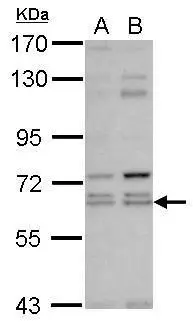
![WB analysis of HeLa cell lysate using GTX83228 KLHL12 antibody [2G2].](https://www.genetex.com/upload/website/prouct_img/normal/GTX83228/GTX83228_20170912_WB_w_23061322_642.webp)
Product group Antibodies
References
C3IP1 antibody [2G2]GTX83228
ApplicationsImmunoFluorescence, Western Blot, ELISA, ImmunoCytoChemistry
ReactivityHuman
TargetKLHL12
- SizePrice
Product group Antibodies

KLHL12 AntibodyLS-C377677
ApplicationsELISA, ImmunoHistoChemistry, ImmunoHistoChemistry Paraffin
ReactivityHuman
TargetKLHL12
- SizePrice
Product group Antibodies
C3IP1 antibody [N1C1]GTX118827
ApplicationsWestern Blot
ReactivityHuman
TargetKLHL12
- SizePrice

Product group Antibodies
Anti-KLHL12 Antibody Picoband(r)A08568-2-CARRIER-FREE
ApplicationsFlow Cytometry, ImmunoFluorescence, Western Blot, ELISA, ImmunoCytoChemistry, ImmunoHistoChemistry
ReactivityHuman, Mouse, Rat
TargetKLHL12
- SizePrice