

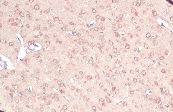
Calnexin antibody [C3], C-term detects Calnexin protein at cell membrane and cytoplasm by immunohistochemical analysis. Sample: Paraffin-embedded rat brain. Calnexin stained by Calnexin antibody [C3], C-term (GTX109669) diluted at 1:165. Antigen Retrieval: Citrate buffer, pH 6.0, 15 min
![Calnexin antibody [C3], C-term detects Calnexin protein at cell membrane and cytoplasm by immunohistochemical analysis. Sample: Paraffin-embedded mouse stomach. Calnexin stained by Calnexin antibody [C3], C-term (GTX109669) diluted at 1:165. Antigen Retrieval: Citrate buffer, pH 6.0, 15 min](https://www.genetex.com/upload/website/prouct_img/normal/GTX109669/GTX109669_44699_20220701_IHC-P_M_22071401_383.webp "Calnexin antibody [C3], C-term detects Calnexin protein at cell membrane and cytoplasm by immunohistochemical analysis. Sample: Paraffin-embedded mouse stomach. Calnexin stained by Calnexin antibody [C3], C-term (GTX109669) diluted at 1:165. Antigen Retrieval: Citrate buffer, pH 6.0, 15 min")
![Calnexin antibody [C3], C-term detects Calnexin protein at endoplasmic reticulum by immunohistochemical analysis. Sample: Paraffin-embedded rat duodenum. Calnexin stained by Calnexin antibody [C3], C-term (GTX109669) diluted at 1:500. Antigen Retrieval: Citrate buffer, pH 6.0, 15 min](https://www.genetex.com/upload/website/prouct_img/normal/GTX109669/GTX109669_43768_20220722_IHC-P_R_22080119_976.webp "Calnexin antibody [C3], C-term detects Calnexin protein at endoplasmic reticulum by immunohistochemical analysis. Sample: Paraffin-embedded rat duodenum. Calnexin stained by Calnexin antibody [C3], C-term (GTX109669) diluted at 1:500. Antigen Retrieval: Citrate buffer, pH 6.0, 15 min")
![Calnexin antibody [C3], C-term detects Calnexin protein at endoplasmic reticulum by immunohistochemical analysis. Sample: Paraffin-embedded mouse duodenum. Calnexin stained by Calnexin antibody [C3], C-term (GTX109669) diluted at 1:500. Antigen Retrieval: Citrate buffer, pH 6.0, 15 min](https://www.genetex.com/upload/website/prouct_img/normal/GTX109669/GTX109669_43768_20220722_IHC-P_M_22080119_426.webp "Calnexin antibody [C3], C-term detects Calnexin protein at endoplasmic reticulum by immunohistochemical analysis. Sample: Paraffin-embedded mouse duodenum. Calnexin stained by Calnexin antibody [C3], C-term (GTX109669) diluted at 1:500. Antigen Retrieval: Citrate buffer, pH 6.0, 15 min")
![Calnexin antibody [C3], C-term detects Calnexin protein at endoplasmic reticulum by immunofluorescent analysis. Sample: HeLa cells were fixed in 4% paraformaldehyde at RT for 15 min. Green: Calnexin stained by Calnexin antibody [C3], C-term (GTX109669) diluted at 1:500. Red: alpha Tubulin, a cytoskeleton marker, stained by alpha Tubulin antibody [GT114] (GTX628802) diluted at 1:1000. Blue: Fluoroshield with DAPI (GTX30920).](https://www.genetex.com/upload/website/prouct_img/normal/GTX109669/GTX109669_44699_20220916_ICC_IF_22102723_612.webp "Calnexin antibody [C3], C-term detects Calnexin protein at endoplasmic reticulum by immunofluorescent analysis. Sample: HeLa cells were fixed in 4% paraformaldehyde at RT for 15 min. Green: Calnexin stained by Calnexin antibody [C3], C-term (GTX109669) diluted at 1:500. Red: alpha Tubulin, a cytoskeleton marker, stained by alpha Tubulin antibody [GT114] (GTX628802) diluted at 1:1000. Blue: Fluoroshield with DAPI (GTX30920).")
![Calnexin antibody [C3], C-term detects Calnexin protein by Western blot analysis. A. 30 μg Neuro2A whole cell lysate/extract B. 30 μg GL261 whole cell lysate/extract C. 30 μg C8D30 whole cell lysate/extract D. 30 μg NIH-3T3 whole cell lysate/extract E. 30 μg BCL-1 whole cell lysate/extract F. 30 μg Raw264.7 whole cell lysate/extract G. 30 μg C2C12 whole cell lysate/extract 7.5 % SDS-PAGE Calnexin antibody [C3], C-term (GTX109669) dilution: 1:10000](https://www.genetex.com/upload/website/prouct_img/normal/GTX109669/GTX109669_41094_WB_M_w_23060500_559.webp "Calnexin antibody [C3], C-term detects Calnexin protein by Western blot analysis. A. 30 μg Neuro2A whole cell lysate/extract B. 30 μg GL261 whole cell lysate/extract C. 30 μg C8D30 whole cell lysate/extract D. 30 μg NIH-3T3 whole cell lysate/extract E. 30 μg BCL-1 whole cell lysate/extract F. 30 μg Raw264.7 whole cell lysate/extract G. 30 μg C2C12 whole cell lysate/extract 7.5 % SDS-PAGE Calnexin antibody [C3], C-term (GTX109669) dilution: 1:10000")
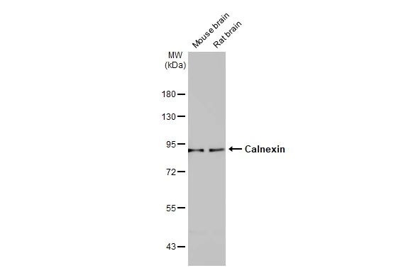
![Various tissue extracts (50 μg) were separated by 7.5% SDS-PAGE, and the membrane was blotted with Calnexin antibody [C3], C-term (GTX109669) diluted at 1:1000. The HRP-conjugated anti-rabbit IgG antibody (GTX213110-01) was used to detect the primary antibody.](https://www.genetex.com/upload/website/prouct_img/normal/GTX109669/GTX109669_43768_20200306_WB_M_R_w_23060500_914.webp "Various tissue extracts (50 μg) were separated by 7.5% SDS-PAGE, and the membrane was blotted with Calnexin antibody [C3], C-term (GTX109669) diluted at 1:1000. The HRP-conjugated anti-rabbit IgG antibody (GTX213110-01) was used to detect the primary antibody.")
![Non-transfected (–) and transfected (+) 293T whole cell extracts (15 μg) were separated by 7.5% SDS-PAGE, and the membrane was blotted with Calnexin antibody [C3], C-term (GTX109669) diluted at 1:20000. The HRP-conjugated anti-rabbit IgG antibody (GTX213110-01) was used to detect the primary antibody.](https://www.genetex.com/upload/website/prouct_img/normal/GTX109669/GTX109669_40450_20160707_WB_shRNA_watermark_w_23060500_509.webp "Non-transfected (–) and transfected (+) 293T whole cell extracts (15 μg) were separated by 7.5% SDS-PAGE, and the membrane was blotted with Calnexin antibody [C3], C-term (GTX109669) diluted at 1:20000. The HRP-conjugated anti-rabbit IgG antibody (GTX213110-01) was used to detect the primary antibody.")
![Non-transfected (–) and transfected (+) 293T whole cell extracts (30 μg) were separated by 7.5% SDS-PAGE, and the membrane was blotted with Calnexin antibody [C3], C-term (GTX109669) diluted at 1:5000. The HRP-conjugated anti-rabbit IgG antibody (GTX213110-01) was used to detect the primary antibody.](https://www.genetex.com/upload/website/prouct_img/normal/GTX109669/GTX109669_43768_20201030_WB_B_w_23060500_521.webp "Non-transfected (–) and transfected (+) 293T whole cell extracts (30 μg) were separated by 7.5% SDS-PAGE, and the membrane was blotted with Calnexin antibody [C3], C-term (GTX109669) diluted at 1:5000. The HRP-conjugated anti-rabbit IgG antibody (GTX213110-01) was used to detect the primary antibody.")
![Various whole cell extracts (30 μg) were separated by 7.5% SDS-PAGE, and the membranes were blotted with Calnexin antibody [C3], C-term (GTX109669) diluted at 1:5000 and competitor's antibody diluted at 1:5000. The HRP-conjugated anti-rabbit IgG antibody (GTX213110-01) was used to detect the primary antibody. *The competitor is not affiliated with GeneTex and does not endorse this product.](https://www.genetex.com/upload/website/prouct_img/normal/GTX109669/GTX109669_43768_20200214_WB_competitor_watermark_w_23060500_471.webp "Various whole cell extracts (30 μg) were separated by 7.5% SDS-PAGE, and the membranes were blotted with Calnexin antibody [C3], C-term (GTX109669) diluted at 1:5000 and competitor's antibody diluted at 1:5000. The HRP-conjugated anti-rabbit IgG antibody (GTX213110-01) was used to detect the primary antibody. *The competitor is not affiliated with GeneTex and does not endorse this product.")
Calnexin antibody [C3], C-term detects Calnexin protein at cell membrane and cytoplasm by immunohistochemical analysis. Sample: Paraffin-embedded rat brain. Calnexin stained by Calnexin antibody [C3], C-term (GTX109669) diluted at 1:165. Antigen Retrieval: Citrate buffer, pH 6.0, 15 min
Calnexin antibody [C3], C-term
GTX109669
ApplicationsImmunoFluorescence, ImmunoPrecipitation, Western Blot, ImmunoCytoChemistry, ImmunoHistoChemistry, ImmunoHistoChemistry Paraffin
Product group Antibodies
ReactivityHuman, Mouse, Rat, Sheep
TargetCANX
Overview
- SupplierGeneTex
- Product NameCalnexin antibody [C3], C-term
- Delivery Days Customer9
- Application Supplier NoteWB: 1:5000-1:20000. ICC/IF: 1:100-1:1000. IHC-P: 1:100-1:1000. IP: 1:500-1:1000. *Optimal dilutions/concentrations should be determined by the researcher.Not tested in other applications.
- ApplicationsImmunoFluorescence, ImmunoPrecipitation, Western Blot, ImmunoCytoChemistry, ImmunoHistoChemistry, ImmunoHistoChemistry Paraffin
- CertificationResearch Use Only
- ClonalityPolyclonal
- Concentration1.2 mg/ml
- ConjugateUnconjugated
- Gene ID821
- Target nameCANX
- Target descriptioncalnexin
- Target synonymsCNX, IP90, P90, calnexin, epididymis secretory sperm binding protein, major histocompatibility complex class I antigen-binding protein p88
- HostRabbit
- IsotypeIgG
- Protein IDP27824
- Protein NameCalnexin
- Scientific DescriptionThis gene encodes a member of the calnexin family of molecular chaperones. The encoded protein is a calcium-binding, endoplasmic reticulum (ER)-associated protein that interacts transiently with newly synthesized N-linked glycoproteins, facilitating protein folding and assembly. It may also play a central role in the quality control of protein folding by retaining incorrectly folded protein subunits within the ER for degradation. Alternatively spliced transcript variants encoding the same protein have been described. [provided by RefSeq]
- ReactivityHuman, Mouse, Rat, Sheep
- Storage Instruction-20°C or -80°C,2°C to 8°C
- UNSPSC12352203
References
- van Wessem KJP, Leenen LPH, Houwert RM, et al. Outcome of severely injured patients in a unique trauma system with 24/7 double trauma surgeon on-call service. Scand J Trauma Resusc Emerg Med. 2023,31(1):60. doi: 10.1186/s13049-023-01122-9Read this paper
- Hsu XR, Wu JE, Wu YY, et al. Exosomal long noncoding RNA MLETA1 promotes tumor progression and metastasis by regulating the miR-186-5p/EGFR and miR-497-5p/IGF1R axes in non-small cell lung cancer. J Exp Clin Cancer Res. 2023,42(1):283. doi: 10.1186/s13046-023-02859-yRead this paper
- Kim M, McDonald EF, Sabusap CMP, et al. Elexacaftor/VX-445-mediated CFTR interactome remodeling reveals differential correction driven by mutation-specific translational dynamics. J Biol Chem. 2023,299(10):105242. doi: 10.1016/j.jbc.2023.105242Read this paper
- Zhao X, Sun Y, Wang Z, et al. Huntingtin exon 1 deletion does not alter the subcellular distribution of huntingtin and gene transcription in mice. Front Cell Neurosci. 2022,16:1021592. doi: 10.3389/fncel.2022.1021592Read this paper
- Gao G, Wang L, Li C. Circ_0006790 carried by bone marrow mesenchymal stem cell-derived exosomes regulates S100A11 DNA methylation through binding to CBX7 in pancreatic ductal adenocarcinoma. Am J Cancer Res. 2022,12(5):1934-1959.Read this paper
- Vultaggio-Poma V, Falzoni S, Chiozzi P, et al. Extracellular ATP is increased by release of ATP-loaded microparticles triggered by nutrient deprivation. Theranostics. 2022,12(2):859-874. doi: 10.7150/thno.66274Read this paper
- Pegoraro A, De Marchi E, Ferracin M, et al. P2X7 promotes metastatic spreading and triggers release of miRNA-containing exosomes and microvesicles from melanoma cells. Cell Death Dis. 2021,12(12):1088. doi: 10.1038/s41419-021-04378-0Read this paper
- Wright MT, Kouba L, Plate L. Thyroglobulin Interactome Profiling Defines Altered Proteostasis Topology Associated With Thyroid Dyshormonogenesis. Mol Cell Proteomics. 2021,20:100008. doi: 10.1074/mcp.RA120.002168Read this paper
- Hu W, Liu Z, Salato V, et al. NOGOB receptor-mediated RAS signaling pathway is a target for suppressing proliferating hemangioma. JCI Insight. 2021,6(3):pii: 142299. doi: 10.1172/jci.insight.142299.Read this paper
- Davies JP, Almasy KM, McDonald EF, et al. Comparative Multiplexed Interactomics of SARS-CoV-2 and Homologous Coronavirus Nonstructural Proteins Identifies Unique and Shared Host-Cell Dependencies. ACS Infect Dis. 2020,6(12):3174-3189. doi: 10.1021/acsinfecdis.0c00500Read this paper
Datasheet
Related products
Product group Antibodies
Anti-CANX Antibody144-63846
ApplicationsImmunoFluorescence, Western Blot, ImmunoHistoChemistry
ReactivityHuman, Mouse, Rat
TargetCANX
- SizePrice

Product group Antibodies
Anti-Calnexin/CANX Antibody Picoband(r)A03372-2-CARRIER-FREE
ApplicationsFlow Cytometry, ImmunoFluorescence, Western Blot, ELISA, ImmunoCytoChemistry, ImmunoHistoChemistry
ReactivityHuman, Mouse
TargetCANX
- SizePrice
![Various whole cell extracts (30 μg) were separated by 7.5% SDS-PAGE, and the membrane was blotted with Calnexin antibody [N3C2], Internal (GTX101676) diluted at 1:5000. The HRP-conjugated anti-rabbit IgG antibody (GTX213110-01) was used to detect the primary antibody.](https://www.genetex.com/upload/website/prouct_img/normal/GTX101676/GTX101676_43643_20190719_WB_w_23060100_608.webp)
Product group Antibodies
References
Calnexin antibody [N3C2], InternalGTX101676
ApplicationsImmunoFluorescence, Western Blot, ImmunoCytoChemistry, ImmunoHistoChemistry, ImmunoHistoChemistry Paraffin
ReactivityHuman, Mouse, Rat
TargetCANX
- SizePrice
Product group Antibodies
References
Calnexin antibodyGTX112886
ApplicationsImmunoFluorescence, Western Blot, ImmunoCytoChemistry, ImmunoHistoChemistry, ImmunoHistoChemistry Paraffin
ReactivityHuman, Mouse, Rat
TargetCANX
- SizePrice
Product group Antibodies
Calnexin antibody [AT18B9]GTX57717
ApplicationsFlow Cytometry, ImmunoFluorescence, Western Blot, ImmunoCytoChemistry
ReactivityHuman
TargetCANX
- SizePrice
![Calnexin antibody [GT1563] detects Calnexin protein at cytoplasm by immunofluorescent analysis. Sample: Neuro2A cells were fixed in 4% paraformaldehyde at RT for 15 min. Green: Calnexin stained by Calnexin antibody [GT1563] (GTX629976) diluted at 1:500. Blue: Fluoroshield with DAPI (GTX30920).](https://www.genetex.com/upload/website/prouct_img/normal/GTX629976/GTX629976_41484_20230512_ICC_IF_M_23060622_501.webp)
Product group Antibodies
References
Calnexin antibody [GT1563]GTX629976
ApplicationsDot Blot, ImmunoFluorescence, ImmunoPrecipitation, Western Blot, ImmunoCytoChemistry
ReactivityHuman, Mouse, Rat
TargetCANX
- SizePrice
![Calnexin antibody [HL1598] detects Calnexin protein at cytoplasm by immunohistochemical analysis. Sample: Paraffin-embedded mouse intestine. Calnexin stained by Calnexin antibody [HL1598] (GTX637077) diluted at 1:100. Antigen Retrieval: Citrate buffer, pH 6.0, 15 min](https://www.genetex.com/upload/website/prouct_img/normal/GTX637077/GTX637077_T-44725_20220722_IHC-P_M_22080119_403.webp)
Product group Antibodies
Calnexin antibody [HL1598]GTX637077
ApplicationsWestern Blot, ImmunoHistoChemistry, ImmunoHistoChemistry Paraffin
ReactivityCanine, Feline, Human, Mouse, Rat
TargetCANX
- SizePrice
Product group Antibodies
CANX Polyclonal AntibodyCAC14640
ApplicationsImmunoFluorescence, Western Blot, ELISA, ImmunoHistoChemistry
TargetCANX
- SizePrice
Product group Antibodies
References
Calnexin Polyclonal AntibodyBS-1693R
ApplicationsFlow Cytometry, ImmunoFluorescence, Western Blot, ELISA, ImmunoCytoChemistry, ImmunoHistoChemistry, ImmunoHistoChemistry Frozen, ImmunoHistoChemistry Paraffin
ReactivityBovine, Chicken, Equine, Human, Mouse, Rabbit, Rat
TargetCANX
- SizePrice
Product group Antibodies
CANX AntibodyCSB-PA001175
ApplicationsImmunoFluorescence, Western Blot, ELISA, ImmunoHistoChemistry
ReactivityHuman, Mouse, Rat
TargetCANX
- SizePrice